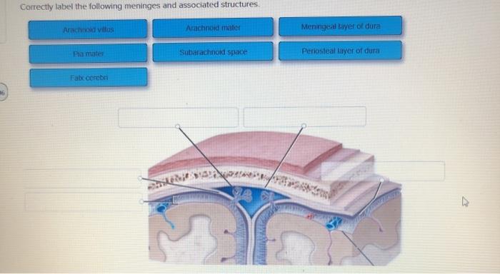
45 correctly label the following meninges and associated structures
Junqueira's Basic Histology Text and Atlas, 14th Edition Web1 H istology is the study of the tissues of the body and how these tissues are arranged to constitute organs. This subject involves all aspects of tissue biology, with the focus on how cells' structure and arrangement optimize functions specific to each organ. › Details › F2012C00424Therapeutic Goods (Medical Devices) Regulations 2002 Jul 04, 2012 · (c) whether the devices are correctly classified under Division 3.1 of Part 3; (d) whether procedures are in place, including a written agreement with the manufacturer of the devices, that require the manufacturer to make available information mentioned in paragraph 41FD (e) or (g) of the Act;
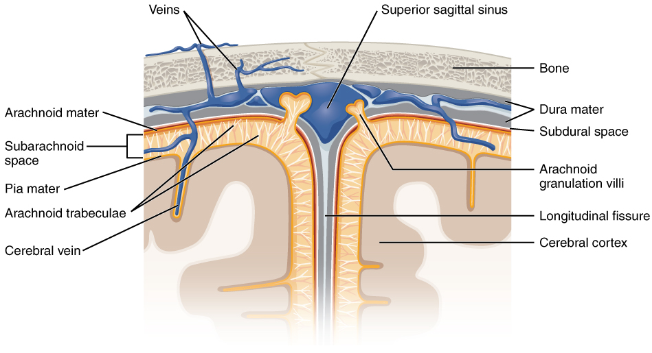
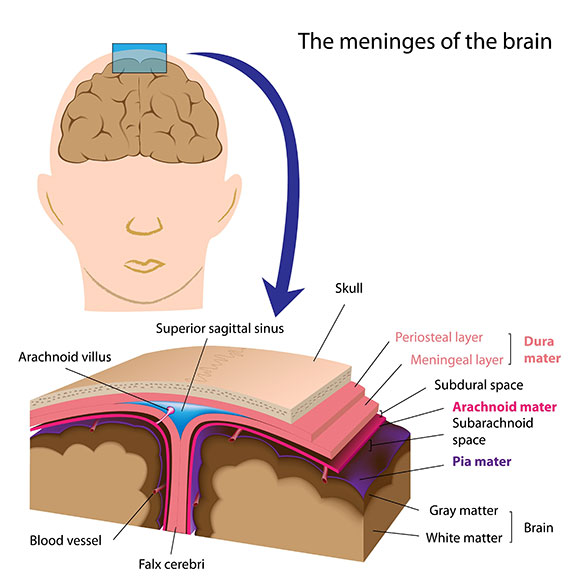
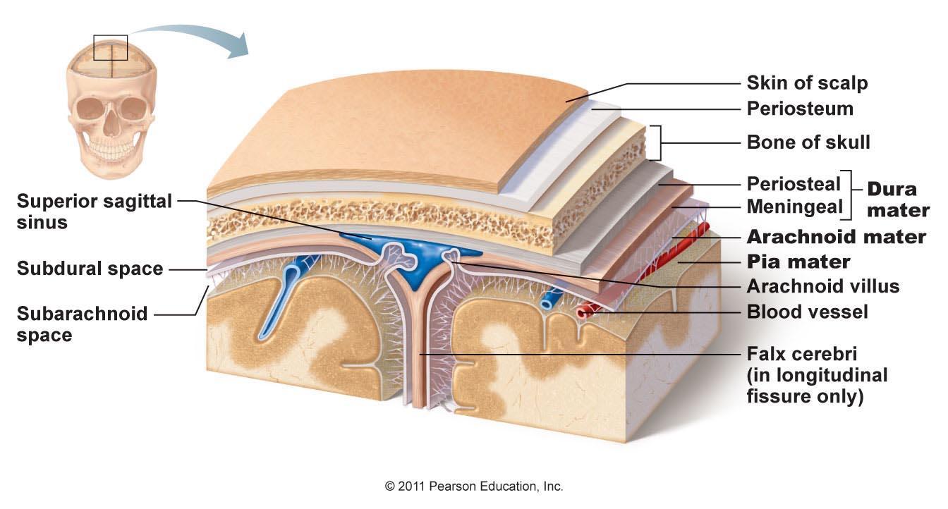
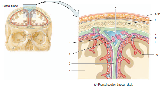
From outermost to innermost, what are the names and the correct order ... Dura mater Arachnoid mater Pia mater From outermost to innermost, Meninges around the brain has three layers : 1. The dura mater 2. the arachnoid mater and 3. the pia mater. The dura mater has two layers : periosteal and meningeal. There is a space between the dura mater and the arachnoid mater, called subdural space. There is also a space between the arachnoid and the pia mater, which is ...

Correctly label the following meninges and associated structures
Meninges: Function and Layers, and Health Problems - ThoughtCo The meninges are composed of three membrane layers known as the dura mater, arachnoid mater, and pia mater. Each layer of the meninges serves a vital role in the proper maintenance and function of the central nervous system. Function Evelyn Bailey The meninges functions primarily to protect and support the central nervous system (CNS). Chapter 14 Question Set Flashcards | Quizlet WebStudy with Quizlet and memorize flashcards containing terms like Label the spinal nerve branches in the figure., Correctly identify and label the structures associated with the rami of the spinal nerves., Correctly identify and label the dermatome(s) represented by the statement(s) associated with them. and more. art-labeling activity: the spinal meninges and associated structures Structure Tone at 444 Hoes Ln Piscataway NJ 08854. Instructors may assign this figure as an art labeling activity using mastering aptm regional anatomy the body is divided into two main regions the axial and appendicular regions. The major region of the brain. The spinal cord and spinal meninges Posting Komentar.
Correctly label the following meninges and associated structures. A & P Unit 4 Flashcards | Quizlet WebCorrectly label the structures and areas associated with a neuron at rest. Label the features of a myelinated axon. The nervous system is involved in most body functions. Match the following definitions or examples with the correct term. The peripheral nervous system is further subdivided into an afferent (sensory) division and an efferent ( _____ ) … nervous system ch10-12 Flashcards | Quizlet Weblabel the internal structures of the cerebrum and other major parts of the brain. cerbrospinal fluid is. clear and liquid . a soldier suffers a brain injury and becomes unable to speak. The damage is likely in. Broca's area. The dura mater is the outer most layer of the meninges. True. Consider left cerebral hemisphere dominance. Drag each label listing a cerebral … Art-labeling Activity: the Limbic System and Associated Structures The Spinal Cord and Spinal Meninges. Brain structures and their functions Art-labeling Activity. It connects areas of the cerebral cortex that are involved in. The limbic system and associated structures Drag the labels to the appropriate location in the figure. The anterior horns are wider than the posterior horns. Correctly Label the Following Meninges and Associated Structures. The Cerebral meninges Are membranes of connective tissue the one that serves as support and that surround the brain and spinal cord. Dura mater arachnoid mater and pia mater. Label the additional cerebral structures on the right side of the figure Label the regions of gray and white matter in the brain Label the components of the cerebral.
CBIO Figures Flashcards | Quizlet Drag each label into the proper location in order to identify the area that would most likely have been affected. Drag each of the given signs and symptoms of nerve damage to the proper position to indicate the nerve most likely affected by the condition. Drag each label to identify the bony passageway through which the given nerve fibers pass. › eudr › 1993/42/2020-12-31Council Directive 93/42/EEC of 14 June 1993 concerning ... Dec 31, 2020 · Article 4 U.K. Free movement, devices intended for special purposes. 1. Member States shall not create any obstacle to the placing on the market or the putting into service within their territory of devices bearing the CE marking provided for in Article 17 which indicate that they have been the subject of an assessment of their conformity in accordance with the provisions of Article 11. Art-labeling Activity: The spinal meninges and associated structures ... 1. Locate major brain structures on a dissected sheep bran, and describe the function of each structure 2. Locate the major spinal cord structures on a dissected sheep al cord, and describe the function of each structure Spe a) Dorsal view... 22) Which of the three spinal meninges is the most superficial? art-labeling activity: the spinal meninges and associated structures The spinal meninges and associated structures. How many pairs of cranial nerves are there. Terms in this set 9 Art-labeling Activity. Tough fibrous outermost layer. Learn vocabulary terms and more with flashcards games and other study tools. The thalamus is the central region of the diencephalon and. The spinal cord and spinal meninges.
A&P2 Lab 13 HW, A&P2 Lab 12 HW, A&P2 Lab 11 HW, A&P2 Lab … WebStudy with Quizlet and memorize flashcards containing terms like Drag the labels onto the diagram to identify the parts of the kidney., Drag the labels onto the diagram to identify the parts of the kidney., What structure is indicated by the red arrow? and more. Chapter 13 Worksheet Flashcards | Quizlet WebCorrectly label the following anatomical features of the spinal cord. Correctly label the following anatomical features of the spinal cord. Drag each label to the appropriate region of the spinal cord. left side top to bottom: - cervical enlargement - lumbosacral enlargement - medullary cone - cauda equina - terminal filum right side top to bottom: - dural sheath - … Correctly label the following meninges of the brain....ask 8 Correctly label the following meninges of the brain. Arachnoid villus Arachnoid mater Subdural space Meningeal layer Pia mater Periosteal layer Dura mater: Subarachnoid space Falx cerebri Dura mater: Reset Zoom Sep 29 2022 | 01:58 PM | Earl Stokes Verified Expert 7 Votes 8464 Answers This is a sample answer. Answered: Describe the basic mechanism by which… | bartleby Label the following structures. Follicles 0 Colloid O Follicular epithelial cells O… A: Endocrine gland is a gland which liberate their secretion into the blood to reach final target as…
Neurophysiological signatures in Alzheimer's disease are ...
Spinal Meninges Anatomy, Diagram & Function | Body Maps - Healthline There are three layers to the meninges: Dura mater: The outermost membrane, this is the thickest of the three layers and has both an outer and inner layer. It is one of the few structures of...
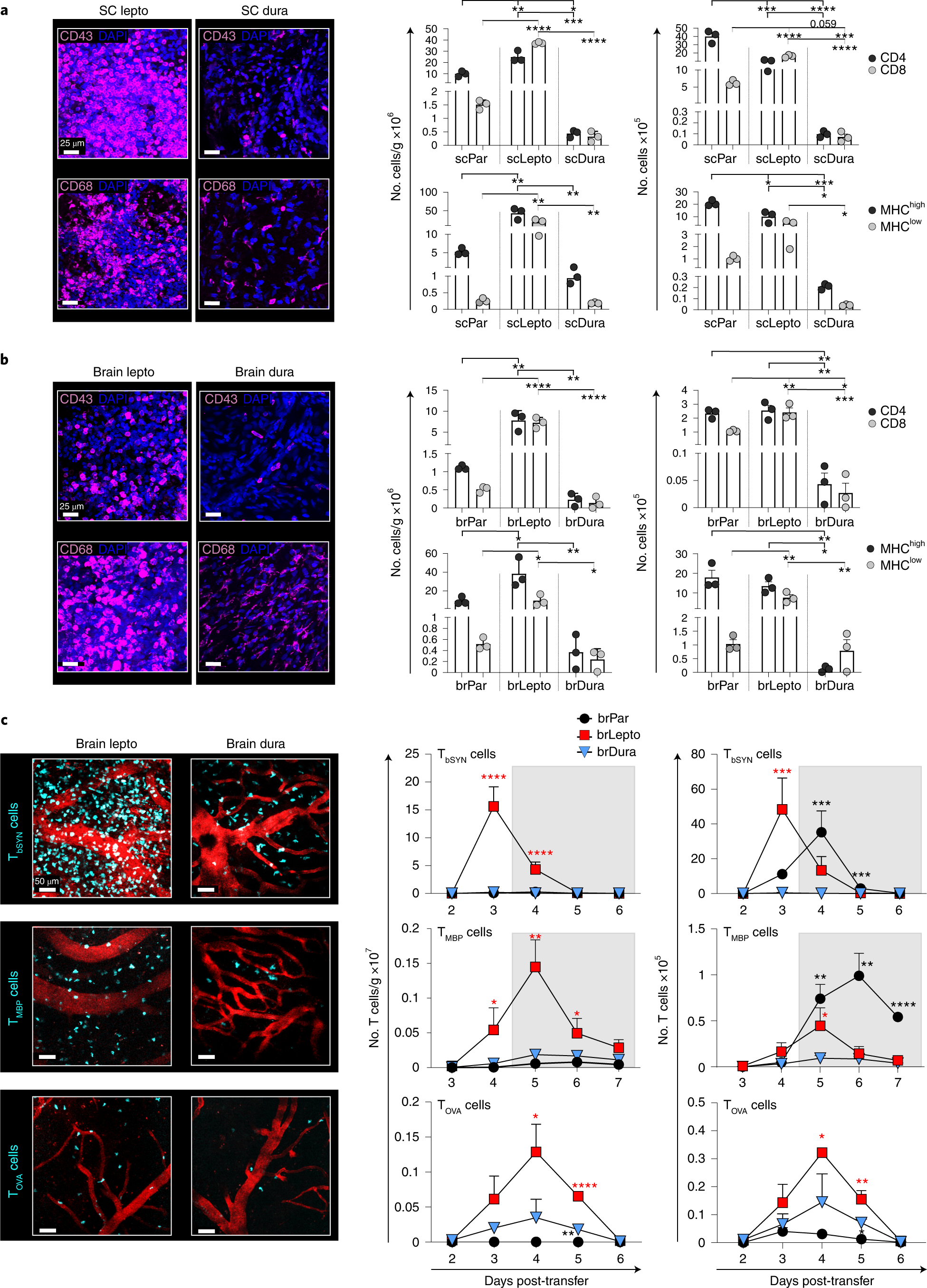
Distinct roles of the meningeal layers in CNS autoimmunity ...
Therapeutic Goods (Medical Devices) Regulations 2002 Web04.07.2012 · (2) If the sponsor of a medical device arranges for a label to be attached or affixed to the device for the purpose of complying with subregulation (1) or for any other purpose (for example, to comply with a labelling requirement under the law of a State or Territory), the label must not in any way adulterate the device or obscure the information …
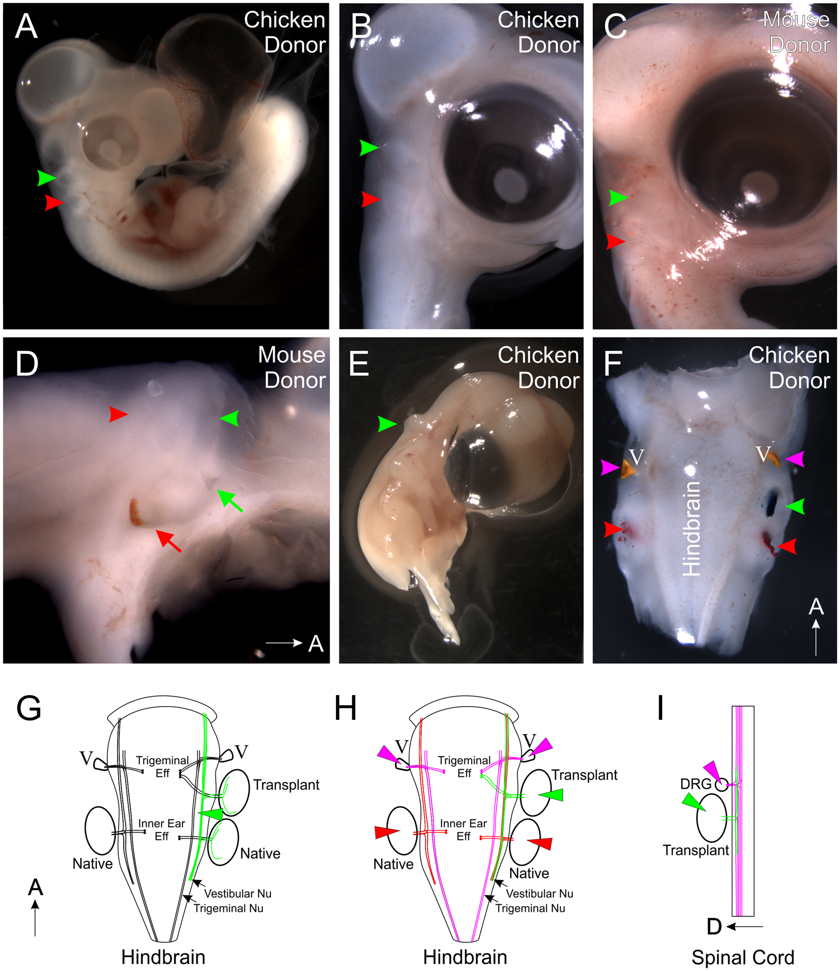
Ear transplantations reveal conservation of inner ear ...
MS Disease Managements - OBSTRUCTION DURING SLEEP (OSA) OVERVIEW ... Read and follow the label, and talk to your doctor or pharmacist if you have questions. for dust mite allergens; pollen is more difficult to avoid because daily activities must be altered to do so; an easy intervention is to keep the windows closed, which is easily accomplished in air- conditioned homes and must be done throughout the year. Use ...

Chapter 13 Question Set Flashcards | Quizlet
Brain & CN Worksheet Flashcards | Quizlet Drag each label into the proper location in order to identify the area that would most likely have been affected. Drag each of the given signs and symptoms of nerve damage to the proper position to indicate the nerve most likely affected by the condition. Assign the appropriate division names of the trigeminal nerve as specified in the figure.

A high-affinity cocaine binding site associated with the ...
Meninges: What They Are & Function - Cleveland Clinic Meninges are three membranes layers that cover and protect your brain and spinal cord (central nervous system). These membranes — the dura mater, arachnoid mater and pia mater — protect and anchor your brain and provide a support system for blood vessels, nerves, lymphatics and the cerebrospinal fluid that surrounds your central nervous system.
Meninges: Dura, arachnoid, pia, meningeal spaces | Kenhub
Meninges and Associated Structures Quiz - PurposeGames.com Meninges and Associated Structures Quiz Science » Image Quiz Meninges and Associated Structures by Mrs. Reid 2,671 plays 14 questions ~ 40 sec 4 too few (you: not rated) Language English Tries 14 [?] Last Played February 22, 2022 - 12:00 am There is a printable worksheet available for download here so you can take the quiz with pen and paper.

Brain meninges labeled Diagram | Quizlet
quizlet.com › 552017541 › nervous-system-ch10-12nervous system ch10-12 Flashcards | Quizlet Study with Quizlet and memorize flashcards containing terms like The motor division is divided further into a somatic motor division serving_____ muscle and a(n)_______ motor division serving_______ muscle, cardiac muscle, and glands., identify each type of neuronal pool, In what type of axon does saltatory conduction occur? and more.

Chapter 14 Worksheet Flashcards | Quizlet
› articles › s41467/020/15906-5Versatile whole-organ/body staining and imaging based on ... Apr 27, 2020 · The following findings from the acquired I-q profiles revealed the structural and chemical characteristics of the delipidated brain. The corresponding d-spacing based on Bragg’s Law ( d = 2π/q ...

Manual sub-segmentation of the cererbellum | medRxiv
Solved 6 Correctly label the following meninges and | Chegg.com Question: 6 Correctly label the following meninges and associated structures Puriosteal layer of dura Arachnoid Villus Arachnoid mater Subarachnoid space Falx corebri Pia mater 0.28 points Meningeal layer of cura eBook Print References Zoom Roset This problem has been solved!
14.2 Blood Flow the meninges and Cerebrospinal Fluid ...
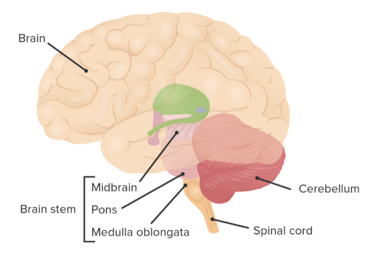
14.3 The Brain and Spinal Cord - Anatomy & Physiology The brain and the spinal cord are the central nervous system, and they represent the main organs of the nervous system. The spinal cord is a single structure, whereas the adult brain is described in terms of four major regions: the cerebrum, the diencephalon, the brain stem, and the cerebellum. A person's conscious experiences are based on ...

25B0CC8B-E772-4BFA-B379-ABCDCE963642.jpeg - Correctly label ...
Chapter 13 Question Set Flashcards | Quizlet Correctly label the following functional regions of the cerebral cortex. Label the regions involved in interpreting and carrying out speech information. Label the diagram with the terms provided to describe the process of neurulation. Cerebrospinal fluid enters the third ventricle of the brain by way of the interventricular foramina.

lab 7 (exercise 14) Flashcards | Quizlet
What midbrain structure is a visual reflex center Superior colliculi of the tectal plate The Meninges of the Brain Correctly label the following meninges and associated structures. The Flow of Cerebrospinal Fluid Place a single word into each sentence to make it correct. Not all terms will be used.
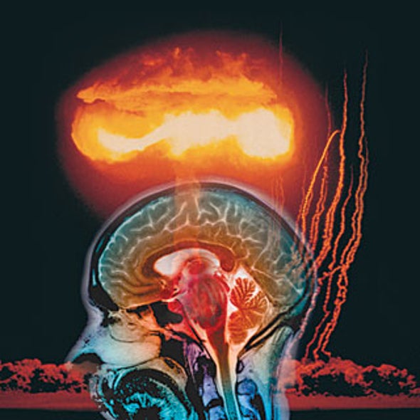
Impact and the Brain - Scientific American
Chapter 13 Question Set Flashcards | Quizlet WebStudy with Quizlet and memorize flashcards containing terms like Identify the cerebral lobes on the left side of the figure. Label the additional cerebral structures on the right side of the figure., Label the regions of gray and white matter in the brain., Label the components of the cerebral nuclei. and more.
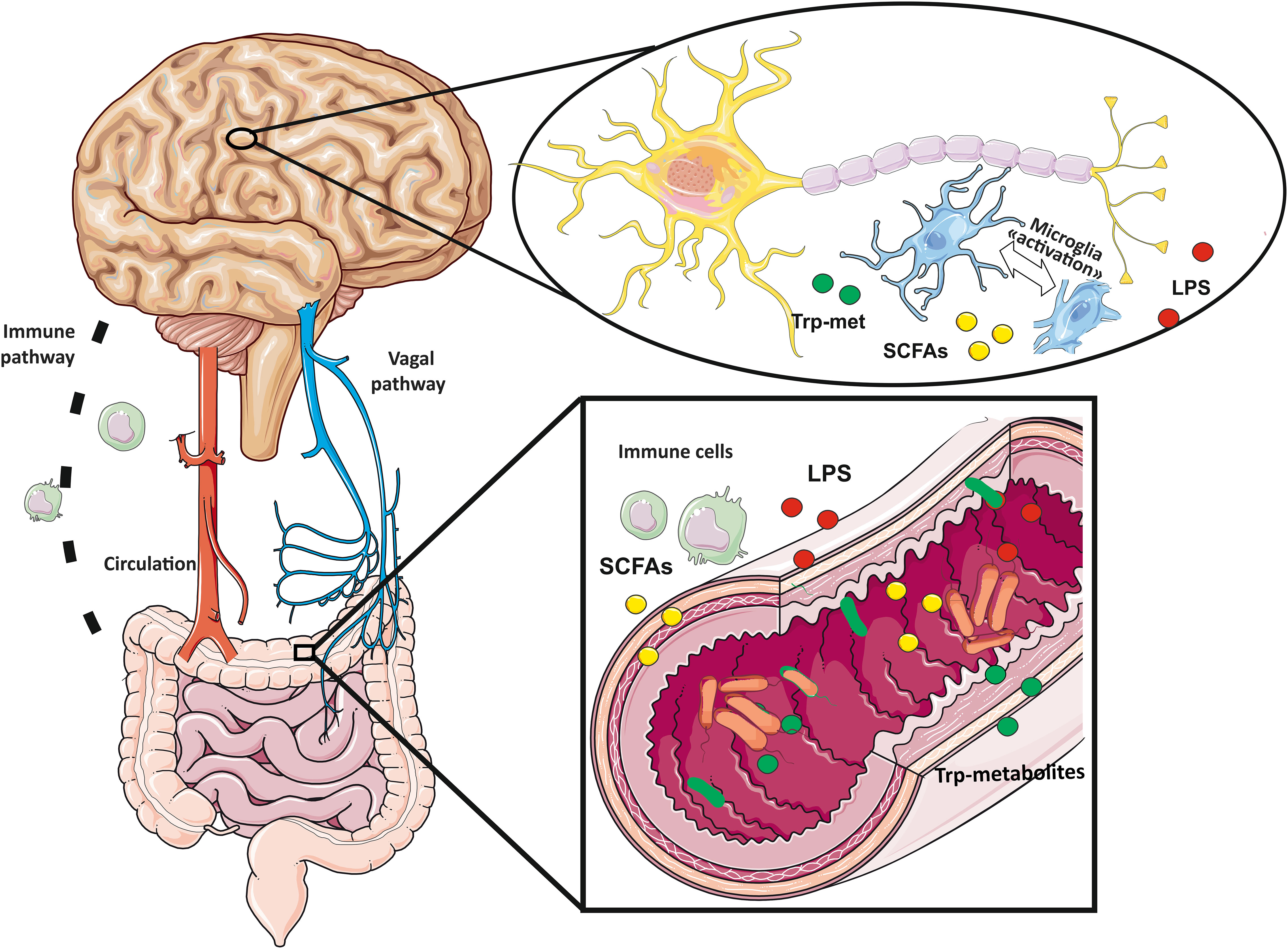
Frontiers | Microglial cells: Sensors for neuronal activity ...
(PDF) Monica Cheesbrough District Laboratory Practice in … WebThe competency of microbiology professionals is obtained through the theoretical knowledge gained in the class and the practical skills and applications that lead to the better understanding and interpretations the subject matter.

Chiari Type I Malformation Associated With Verbal Fluency ...
EUR-Lex - 01993L0042-20071011 - EN - EUR-Lex - Europa Web11.10.2007 · Where a Member State ascertains that the devices referred to in Article 4 (1) and (2) second indent, when correctly installed, maintained and used for their intended purpose, may compromise the health and/or safety of patients, users or, where applicable, other persons, it shall take all appropriate interim measures to withdraw such devices …
Meninges: Anatomy | Concise Medical Knowledge
quizlet.com › 582006615 › chapter-13-worksheet-flashChapter 13 Worksheet Flashcards | Quizlet Correctly label the following anatomical features of the spinal cord. Drag each label to the appropriate region of the spinal cord. left side top to bottom: - cervical enlargement - lumbosacral enlargement - medullary cone - cauda equina - terminal filum right side top to bottom: - dural sheath - subarachnoid space
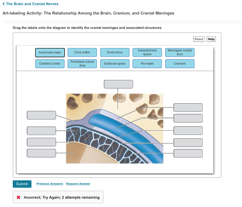
Solved K The Brain and Cranial Nerves Art-labeling Activity ...
quizlet.com › 538793548 › a-p-unit-4-flash-cardsA & P Unit 4 Flashcards | Quizlet Correctly identify and label the dermatome(s) represented by the statement(s) associated with them. Correctly identify the function of each structure that comprises a tendon reflex by dragging the appropriate label into place.
What four structures protect the central nervous system ...
Chapter 14 Question Set - Quizzes Studymoose Correctly identify and label the structures associated with the rami of the spinal nerves. ... represented by the statement(s) associated with them. answer. question. Correctly identify the function of each structure that comprises a tendon reflex by dragging the appropriate label into place. ... Correctly label the following anatomical ...

Area Postrema Cell Types that Mediate Nausea-Associated ...
Solved Correctly label the following meninges and associated - Chegg Question: Correctly label the following meninges and associated structures Arachnoid valus Arachnoid maler Meningeal layer of dura Pia mater Subarachnoid space Periosteal layer of dura Fabcccrobni This problem has been solved! You'll get a detailed solution from a subject matter expert that helps you learn core concepts. See Answer
:max_bytes(150000):strip_icc()/anatomy-of-the-brain--meninges--hypothalamus-and-anterior-pituitary--1134486874-240d1e77d3364d5b8572be4b44a43666.jpg)
Meninges: Function and Layers, and Health Problems
eur-lex.europa.eu › legal-content › ENEUR-Lex - 01993L0042-20071011 - EN - EUR-Lex - Europa Oct 11, 2007 · Whereas it is necessary, essentially for the purpose of the conformity assessment procedures, to group the devices into four product classes; whereas the classification rules are based on the vulnerability of the human body taking account of the potential risks associated with the technical design and manufacture of the devices; whereas the ...
The Spinal Cord
Neuroanatomy, Cranial Meninges - StatPearls - NCBI Bookshelf The brain and spinal cord are enveloped within three layers of membrane collectively known as the meninges, with the cranial meninges specifically referring to the section that covers the brain. From superficial to deep, the three layers are the dura, arachnoid, and pia—the term "mater," Latin for mother, often follows these names (i.e., dura mater, arachnoid mater, pia mater).[1] The ...
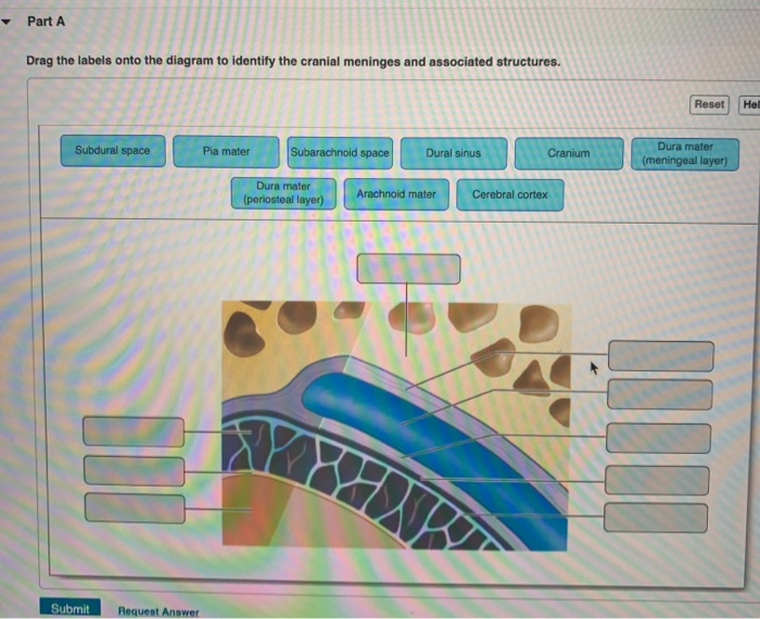
Solved Part A Drag the labels onto the diagram to identify ...
Meninges: Structure and Functions - Exploring your mind The three meninges are the dura mater, the arachnoid mater, and the pia mater. The last two comprise the leptomeninges. The dura mater is also called the pachymeninx. The main function of the meninges is to protect the brain. T his is a very vulnerable organ that needs special protection. No other organ needs it, at least not in the same way.
Nervous System: Anatomy, Structure, and Classification ...
The Meninges - Dura - Arachnoid - Pia - TeachMeAnatomy The meninges refer to the membranous coverings of the brain and spinal cord. There are three layers of meninges, known as the dura mater, arachnoid mater and pia mater. These coverings have two major functions: Provide a supportive framework for the cerebral and cranial vasculature.

human skeleton - The lower jaw | Britannica
Meninges: Dura, arachnoid, pia, meningeal spaces | Kenhub Meninges of the brain The meninges are the three membranes that envelop the brain and spinal cord and separate them from the walls of their bony cases ( skull and vertebral column ). Based on their location, meninges are referred to as the cranial meninges which envelop the brain, and spinal meninges which envelop the spinal cord.
From outermost to innermost, what are the names and the ...
Chapter 14 Worksheet Flashcards | Quizlet Correctly label the following meninges of the brain. Place a single word into each sentence to make it correct, then place each sentence into a logical paragraph order describing the flow of cerebrospinal fluid. 1. CSF is secreted into each LATERAL ventricle by choroid plexus and flows into the third ventricle where more is added. 2.

Back Surgery That Does Not Relieve Pain
art-labeling activity: the spinal meninges and associated structures Structure Tone at 444 Hoes Ln Piscataway NJ 08854. Instructors may assign this figure as an art labeling activity using mastering aptm regional anatomy the body is divided into two main regions the axial and appendicular regions. The major region of the brain. The spinal cord and spinal meninges Posting Komentar.
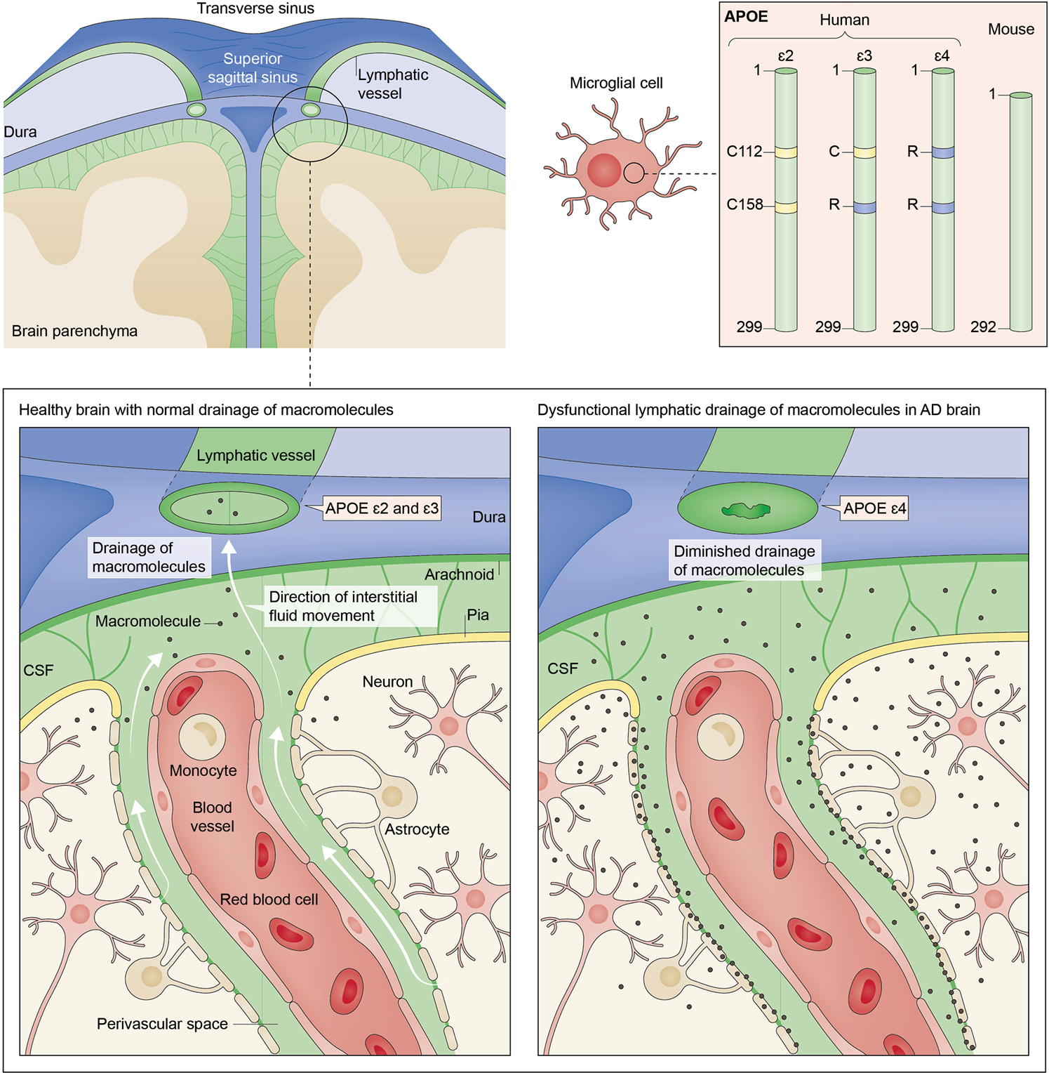
Apolipoprotein E4 and meningeal lymphatics in Alzheimer ...
Chapter 14 Question Set Flashcards | Quizlet WebStudy with Quizlet and memorize flashcards containing terms like Label the spinal nerve branches in the figure., Correctly identify and label the structures associated with the rami of the spinal nerves., Correctly identify and label the dermatome(s) represented by the statement(s) associated with them. and more.
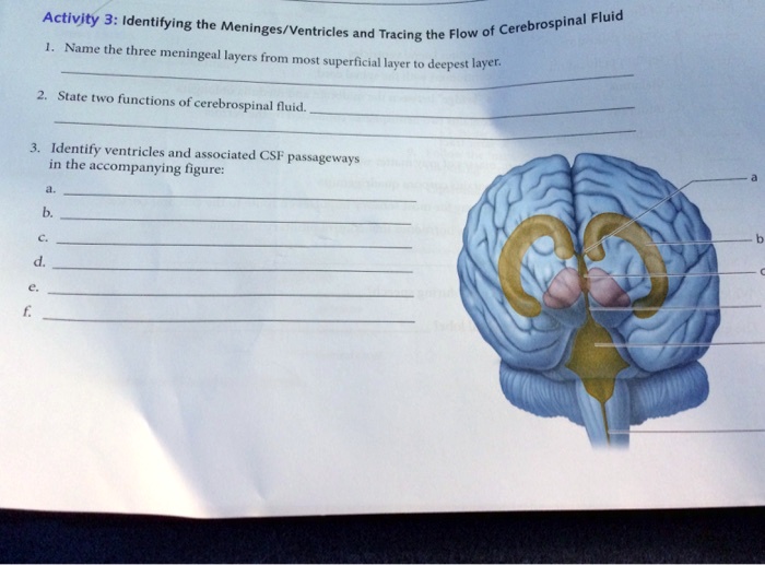
SOLVED: Activity 3: Identifying the Cerebrospinal Fluid ...
Meninges: Function and Layers, and Health Problems - ThoughtCo The meninges are composed of three membrane layers known as the dura mater, arachnoid mater, and pia mater. Each layer of the meninges serves a vital role in the proper maintenance and function of the central nervous system. Function Evelyn Bailey The meninges functions primarily to protect and support the central nervous system (CNS).

A&P 1 final Flashcards | Quizlet

Chapter 13 and 14 Flashcards | Quizlet
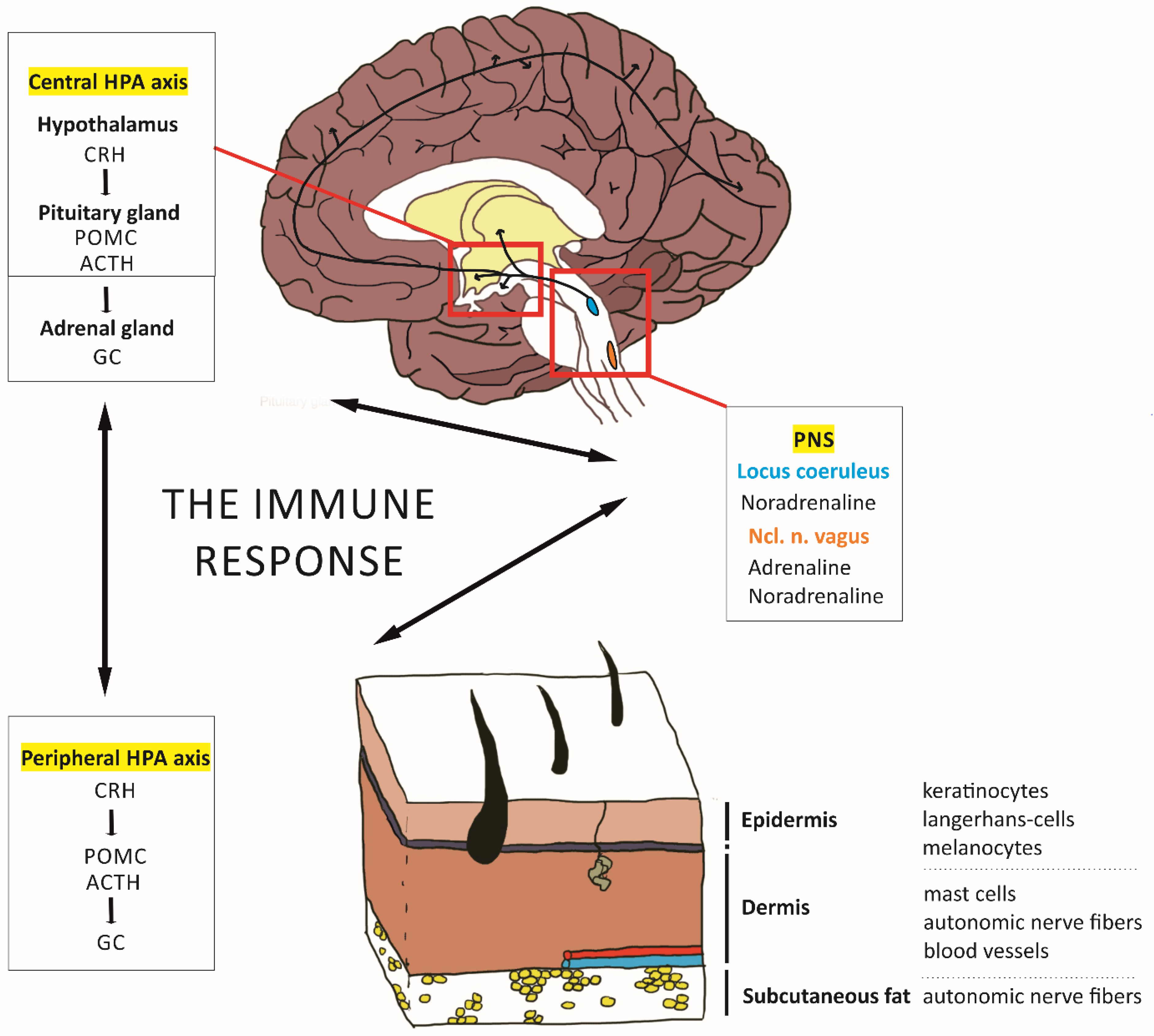
IJMS | Free Full-Text | Probing the Skin–Brain Axis ...

Meninges and Associated Structures Diagram | Quizlet

DB is associated with increased phagocytic activity but not ...
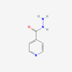
Isoniazid | C6H7N3O - PubChem
Solved Correctly label the following meninges and associated ...

Reassessing Human Adipose Tissue | NEJM

Fluid transport in the brain | Physiological Reviews
Solved] Describe the follow dorsal brain structures below ...
Anatomy Exam 2 Flashcards - Easy Notecards
A deep learning algorithm for 3D cell detection in whole ...

Solved heducation.com Brain and Spinal Cord Correctly label ...
Brain | Definition, Parts, Functions, & Facts | Britannica
Spinal Cord Quiz: Cross-Sectional Anatomy | GetBodySmart
Veins of the brain: Anatomy and clinical notes | Kenhub

Brain endothelial STING1 activation by Plasmodium-sequestered ...
Post a Comment for "45 correctly label the following meninges and associated structures"