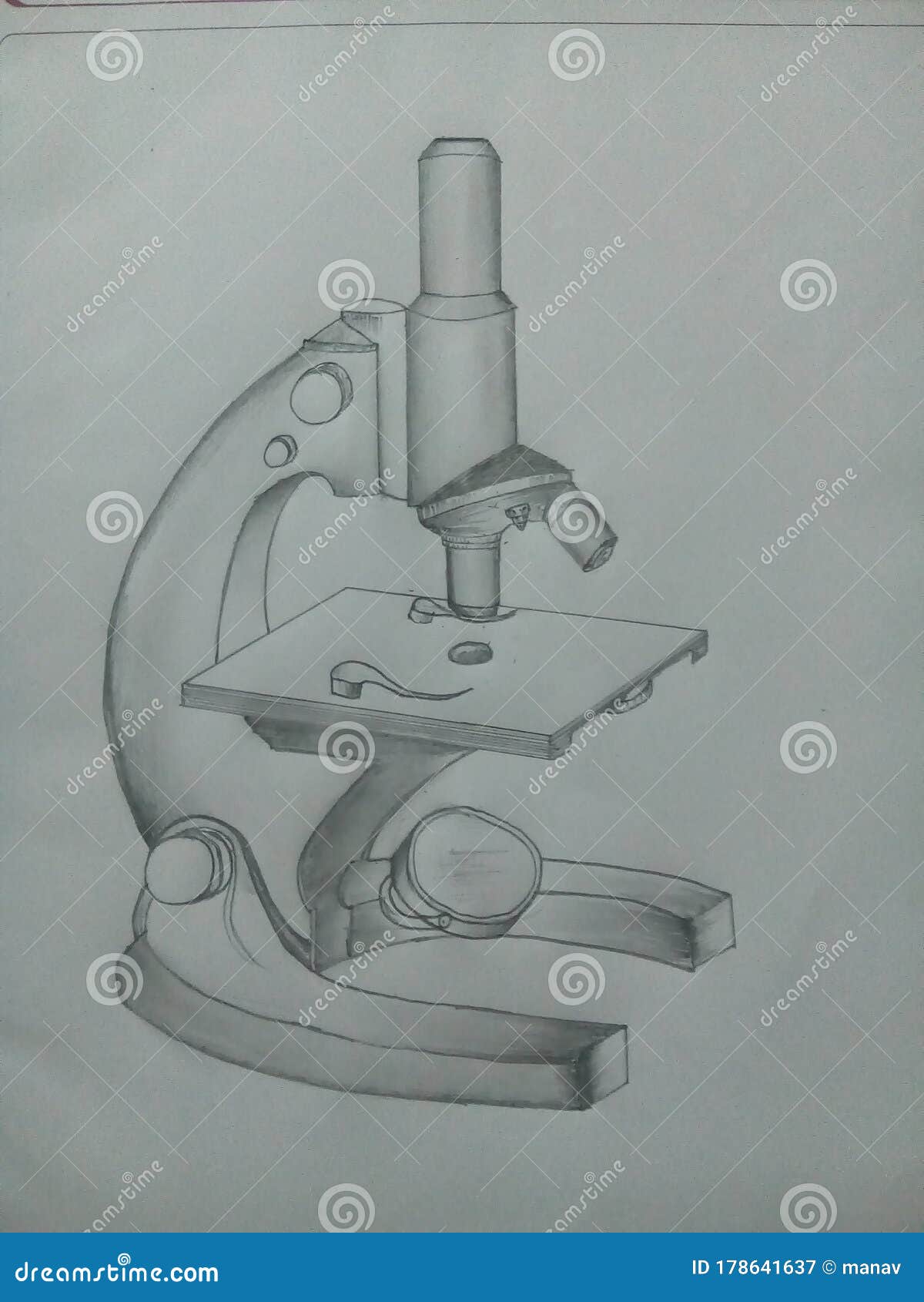
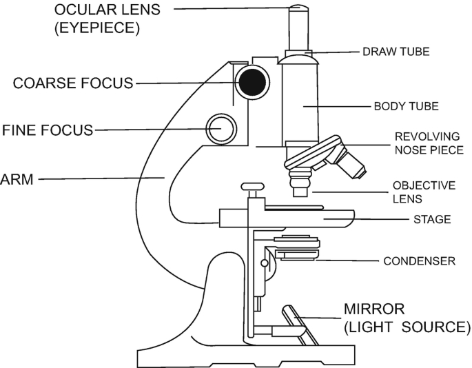
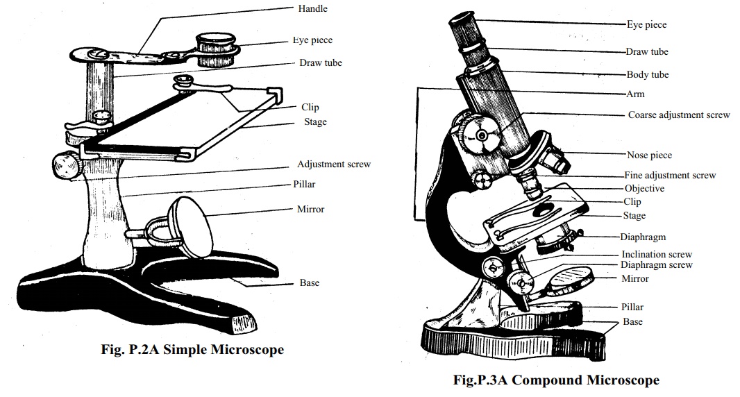
39 parts of compound microscope drawing
Microscope: Types of Microscope, Parts, Uses, Diagram - Embibe A compound microscope is defined as a microscope with a high resolution. It uses two sets of lenses, providing a \(2\)-dimensional image of the sample. The term compound refers to the usage of more than one lens in the microscope. Also, the compound microscope is one of the types of optical microscopes. How to Draw a Microscope - Realistic Microscope Drawing Tutorial Step 3: Outline the Arm Frame. We are now going to draw the arm that the microscope uses to swivel back and forth. Attached to the right side of the head from the previous step, draw two curving lines. You can then make this three-dimensional by drawing two smaller curved lines in the way you can see below.
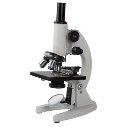
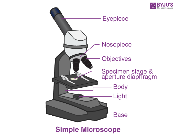
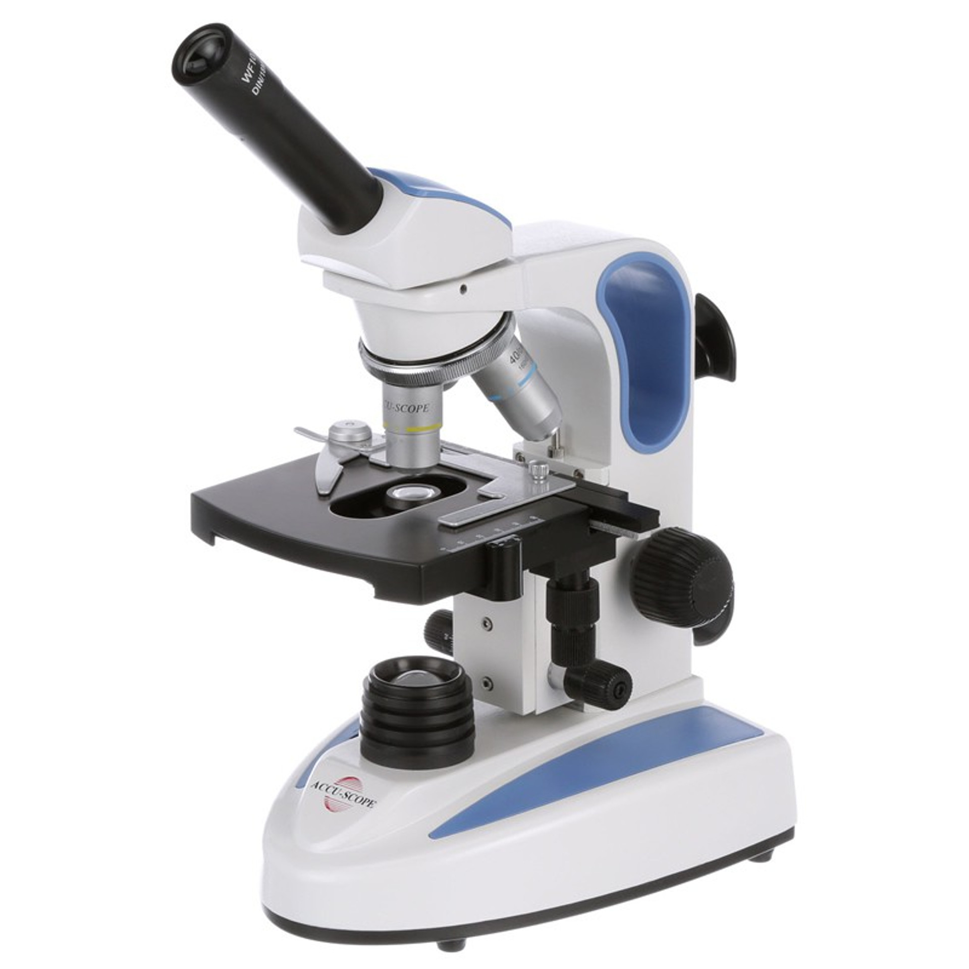
Compound Microscope: Definition, Diagram, Parts, Uses ... - BYJUS The parts of a compound microscope can be classified into two: Non-optical parts Optical parts Non-optical parts Base The base is also known as the foot which is either U or horseshoe-shaped. It is a metallic structure that supports the entire microscope. Pillar The connection between the base and the arm are possible through the pillar. Arm
Parts of compound microscope drawing
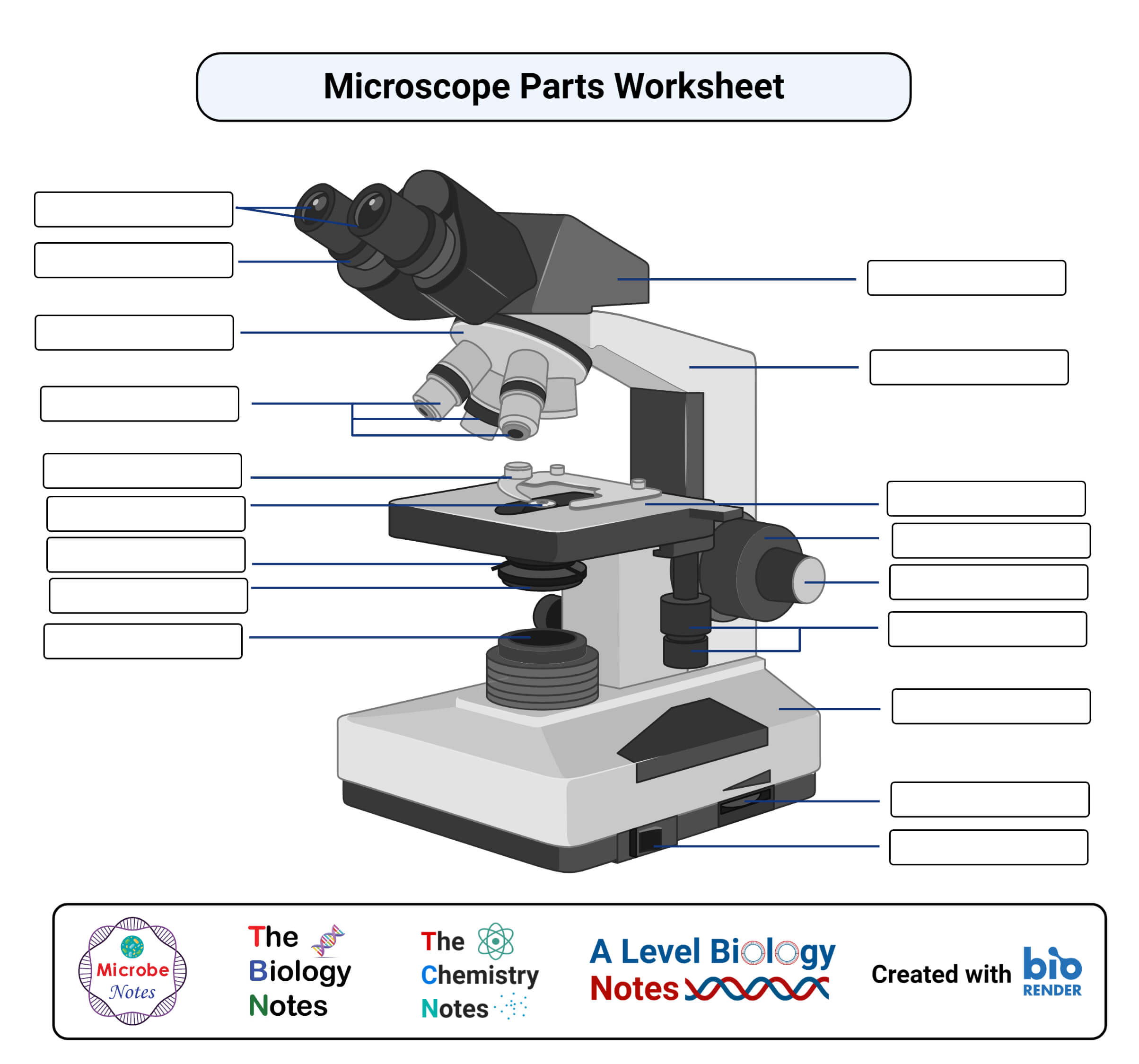
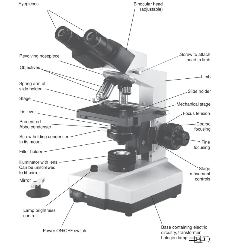
Parts of a Compound Microscope (And their Functions) - Scope Detective List of Microscope Parts and their Functions. 1. Ocular Tubes (Monocular, Binocular & Trinocular) The ocular tubes, are to tubes that lead from the head of the microscope out to your eyes. On the end of the ocular tubes are usually interchangeable eyepieces (commonly 10X and 20X) that increase magnification. Compound Microscope Parts Head/Body houses the optical parts in the upper part of the microscope. Base of the microscope supports the microscope and houses the illuminator. Arm connects to the base and supports the microscope head. It is also used to carry the microscope. When carrying a compound microscope always take care to lift it by both the arm and base ... Parts of a microscope with functions and labeled diagram - Microbe Notes Figure: Diagram of parts of a microscope. There are three structural parts of the microscope i.e. head, base, and arm. Head - This is also known as the body. It carries the optical parts in the upper part of the microscope. Base - It acts as microscopes support. It also carries microscopic illuminators.
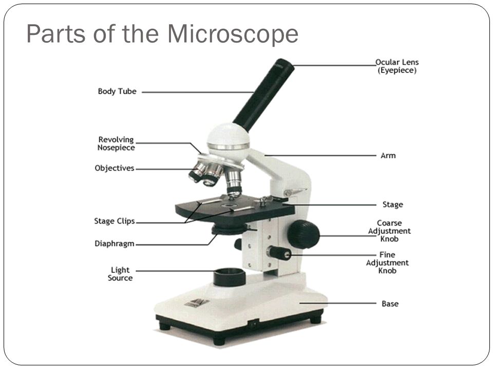
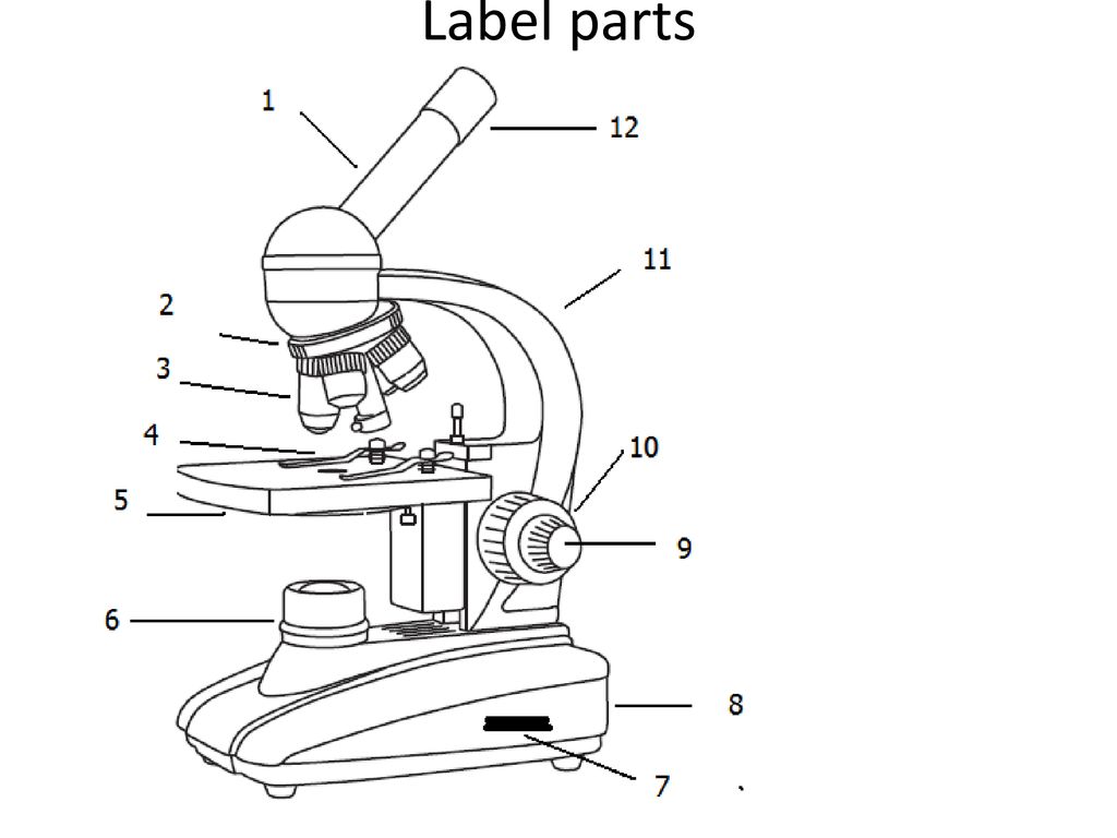
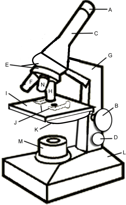
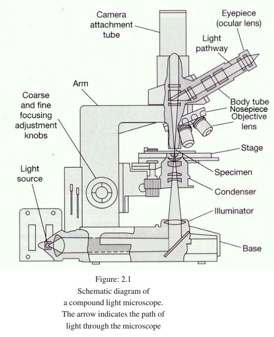
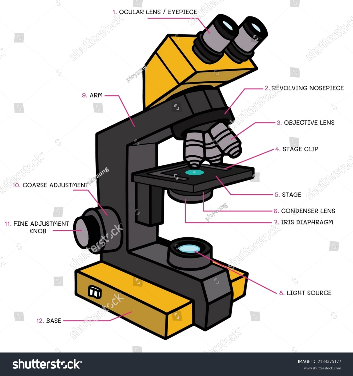
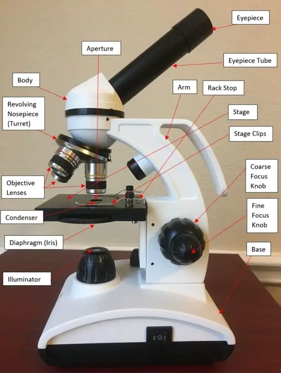
Parts of compound microscope drawing. PDF AN INTRODUCTION TO THE COMPOUND MICROSCOPE - Rowan University **When carrying the microscope, place one hand on the base and the other hand around the arm. **DO NOT PLACE THE MICROSCOPE IN AN UPSIDE DOWN POSITION. PIECES WILL FALL OUT. **Keep microscope away from the edge of the bench, particularly when not in use. **Make sure power cords are out of the way. **Never force the microscope parts to work. Compound Microscope: Parts of Compound Microscope - BYJUS (A) Mechanical Parts of a Compound Microscope 1. Foot or base It is a U-shaped structure and supports the entire weight of the compound microscope. 2. Pillar It is a vertical projection. This stands by resting on the base and supports the stage. 3. Arm The entire microscope is handled by a strong and curved structure known as the arm. 4. Stage Working Principle and Parts of a Compound Microscope (with Diagrams) It has a series of two lenses; (i) the objective lens close to the object to be observed and (ii) the ocular lens or eyepiece, through which the image is viewed by eye. Light from a light source (mirror or electric lamp) passes through a thin transparent object (Figure 4.4). The objective lens produces a magnified 'real image' first image) of the object. Compound Microscope - Diagram (Parts labelled), Principle and Uses What are the 13 parts of a microscope? 1. Eyepiece 2. Eyepiece Tube 3. Objective Lens 4. Stage 5. Stage Clips 6. Nosepiece 7. Fine and Coarse Focus knobs 8. Illuminator 9. Aperture 10. Iris Diaphragm 11. Condenser 12. Condenser Focus Knob 13. The Rack stop Q 5. What are the 11 parts of a compound microscope?
Chapter 5 The Integumentary System – Anatomy and Physiology ... Sketch the skin area with eccrine sweat glands as seen in the microscope, observed at low and high magnification. Exercise 6 Axillaries of Skin – Nail. Required Materials. Compound microscope; Slide of developing nail; Procedure. Obtain a slide of nail, place it on the stage of the microscope and scan it to view all the parts of the nail ... Simple Microscope - Diagram (Parts labelled), Principle, Formula and Uses Simple microscope is a magnification apparatus that uses a combination of double convex lens to form an enlarged, erect image of a specimen. The working principle of a simple microscope is that when a lens is held close to the eye, a virtual, magnified and erect image of a specimen is formed at the least possible distance from which a human eye ... 16 Parts of a Compound Microscope: Diagrams and Video The 16 core parts of a compound microscope are: Head (Body) Arm Base Eyepiece Eyepiece tube Objective lenses Revolving Nosepiece (Turret) Rack stop Coarse adjustment knobs Fine adjustment knobs Stage Stage clips Aperture Illuminator Condenser Diaphragm How to draw compound of Microscope easily - step by step I will show you " How to draw compound of microscope easily - step by step "Please watch carefully and try this okay.Thanks for watching.....#microscopedrawi...
PDF Parts of a Microscope Printables - Homeschool Creations The lenses in a microscope make items appear smaller. How many parts of a microscope can you identify? Can you show the arm, stage, eyepiece, head, objective lens, illuminator, nosepiece, and stage clips? Where is the safest place to hold or carry a microscope? Which part of the microscope holds the specimen slide in place? Why do we use ... Microscope Parts and Functions Here are the important compound microscope parts... Eyepiece: The lens the viewer looks through to see the specimen. The eyepiece usually contains a 10X or 15X power lens. Diopter Adjustment: Useful as a means to change focus on one eyepiece so as to correct for any difference in vision between your two eyes. parts of a microscope diagram Microscope parts science compound knob adjustment coarse labeled diagram biology microscopes light label name tools lab functions does structure 6th. Plant phloem vascular cells anatomy parenchyma primary cambium sclereids fibers tissue sieve elements conducting differentiated procambium initiated secondary called grkraj. Compound Microscope Parts: - cas.miamioh.edu Compound Microscope Parts: To Diagram/ Self-Test. Eyepiece; Objectives; Fine Adjustment Knob ... Coarse Adjustment Knob; Aperture; Arm; Light Source. Return To Top Of Page. Microscopy Main Page. Microscope Parts. Magnification. Microscope Types. Using Microscopes. Resolution. Microscope History. Microscope Care. Who uses microscopes? Compound ...
Parts of Compound Microscope (With Diagram) - Biology Discussion A compound microscope consists of three main parts: 1. Objective Lens 2. Eye Piece 3. Condensers. Part # 1. Objective Lens: The chief characteristics of the objective lens are: (a) To gather the light rays coming any point to the object,
Microscopy - Northern Arizona University Microscope Drawings. When drawing what you see under the microscope, follow the format shown below. It is important to include a figure label and a subject title above the image. The species name (and common name if there is one) and the magnification at which you were viewing the object should be written below the image.
Compound Microscope- Definition, Labeled Diagram, Principle ... Apr 03, 2022 · Parts of a Compound Microscope. Eyepiece And Body Tube. The eyepiece is the lens through which the viewer looks to see the specimen. It usually contains a 10X or 15X power lens. The body tube connects the eyepiece to the objective lenses. Objectives and Stage Clips. Objective Lenses are one of the most important parts of a Compound Microscope.
Compound Microscope Parts, Function, & Diagram | What is a Compound ... There are many functioning parts to the compound light microscope Head/Body The first part of the compound light microscope is the head. This is the top portion of the compound microscope that...
Diagram of a Compound Microscope - Biology Discussion The size of objects viewed under the compound microscope can be accurately determined using a micrometer. The latter consists of two scales, the eyepiece scale, (also called 'graticule' or 'ocular') and the stage micrometer scale. The eyepiece scale is calibrated with the help of stage micrometer and the former is then used for measurements.
Poly(methyl methacrylate) - Wikipedia Poly(methyl methacrylate) (PMMA) belongs to a group of materials called engineering plastics.It is a transparent thermoplastic.PMMA is also known as acrylic, acrylic glass, as well as by the trade names and brands Crylux, Plexiglas, Acrylite, Astariglas, Lucite, Perclax, and Perspex, among several others ().
Word roots: The web’s largest word root and prefix directory microscope - a device used to see tiny things; periscope - a seeing instrument on a submarine; telescope - a device used to see over a distance. scrib, script: write, written: Latin: inscribe - to write letters or words on a surface; scribe - a person who writes out documents; describe - to represent with words or pictures. se: apart: Latin
Compound Microscope Drawing With Parts and Functions - NotesHippo Compound Microscope Drawing With Parts and Functions Objectives The image of the specimen first passes through the objective. Objectives with magnifying powers 4X, 10X, 40X and 100X are commonly used. The magnifying power is marked on the lens and is usually colour-coded for easy identification. Mechanical stage
Compound Microscope Parts Diagram | Quizlet Start studying Compound Microscope Parts. Learn vocabulary, terms, and more with flashcards, games, and other study tools.
Compound Microscope Parts - Labeled Diagram and their Functions Optical components of a compound microscope Eyepiece. The eyepiece (or ocular lens) is the lens part at the top of a microscope that the viewer looks through. Eyepiece tube. The eyepiece tube carries the eyepiece lens. It holds the eyepiece in the right place that aligns... Objective lenses. ...
Compound Microscope Parts, Functions, and Labeled Diagram Parts of a Compound Microscope Eyepiece (ocular lens) with or without Pointer: The part that is looked through at the top of the compound microscope. Monocular or Binocular Head: Structural support that holds & connects the eyepieces to the objective lenses. Arm: Supports the microscope head and ...
What is Compound Microscope? - Diagram, Function, Advantages The Compound Microscope is a special kind of microscope that features two sets of high resolutions lenses, that are capable of providing a 2-dimensional image of the sample. And because of the presence of two lenses in this microscope, it is referred to as a compound microscope. Moreover, based on the purpose of these microscopes, they are ...
Parts of Stereo Microscope (Dissecting microscope) - labeled diagram ... Stereo microscopes (also called Dissecting microscope) are branched out from other light microscopes for the application of viewing "3D" objects. These include substantial specimens, such as insects, feathers, leaves, rocks, sand grains, gems, coins, and stamps, etc. Functionally, a stereo microscope is like a powerful magnifying glass.
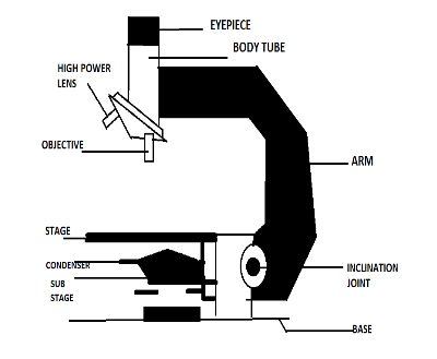
Parts of the Compound Microscope Diagram | Quizlet The Mechanical Parts Base Arm Draw Tube Body Tube Adjusting Knobs Dust Shield Revolving Nosepiece Inclination Joint Stage Iris Diaphragm Pillar Illuminating Parts Condenser Mirror Magnifying Parts Eyepiece or Ocular Objectives Component Parts of the Watson Service Microscope (c. 1950) Eyepiece Body Tube Coarse-Adjustment Fine-Adjustment Limb Joint
Parts of a Compound Microscope and Their Functions - NotesHippo Compound microscope mechanical parts (Microscope Diagram: 2) include base or foot, pillar, arm, inclination joint, stage, clips, diaphragm, body tube, nose piece, coarse adjustment knob and fine adjustment knob. Base: It's the horseshoe-shaped base structure of microscope. All of the other components of the compound microscope are supported by it.
Parts of a microscope with functions and labeled diagram - Microbe Notes Figure: Diagram of parts of a microscope. There are three structural parts of the microscope i.e. head, base, and arm. Head - This is also known as the body. It carries the optical parts in the upper part of the microscope. Base - It acts as microscopes support. It also carries microscopic illuminators.
Compound Microscope Parts Head/Body houses the optical parts in the upper part of the microscope. Base of the microscope supports the microscope and houses the illuminator. Arm connects to the base and supports the microscope head. It is also used to carry the microscope. When carrying a compound microscope always take care to lift it by both the arm and base ...
Parts of a Compound Microscope (And their Functions) - Scope Detective List of Microscope Parts and their Functions. 1. Ocular Tubes (Monocular, Binocular & Trinocular) The ocular tubes, are to tubes that lead from the head of the microscope out to your eyes. On the end of the ocular tubes are usually interchangeable eyepieces (commonly 10X and 20X) that increase magnification.










Post a Comment for "39 parts of compound microscope drawing"