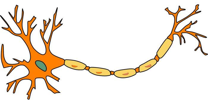
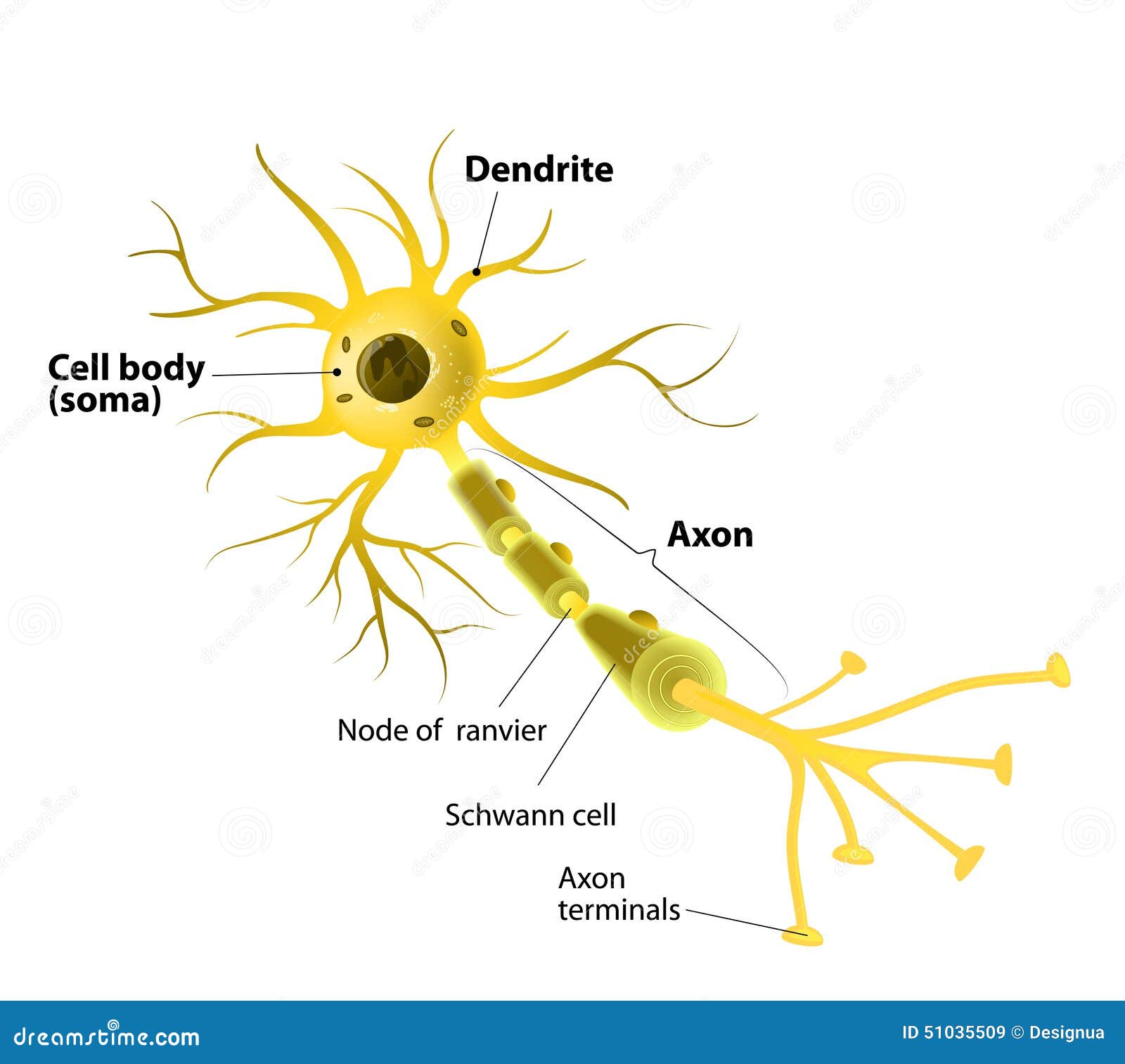
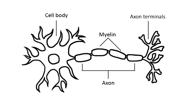
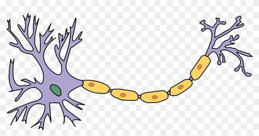
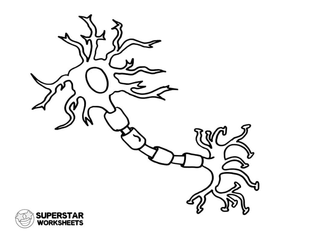
43 diagram of neuron with labels
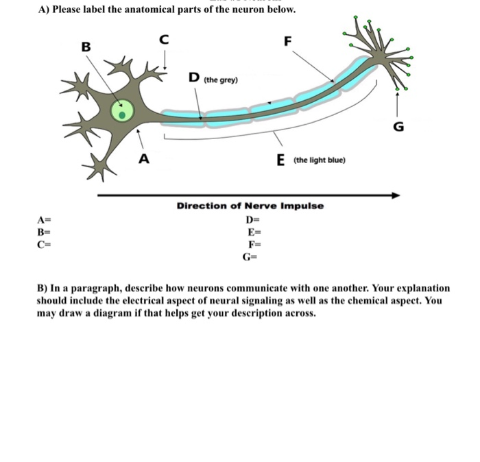
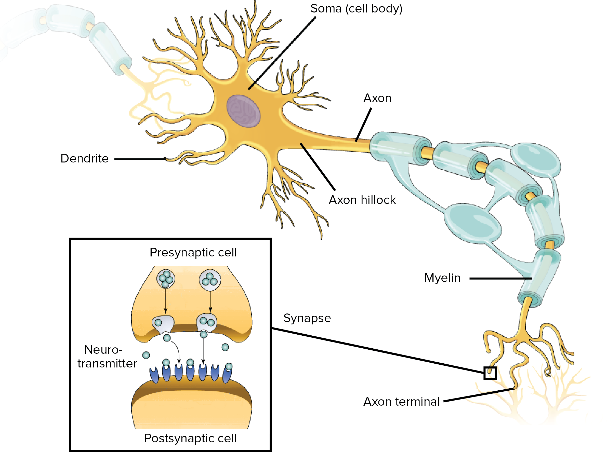
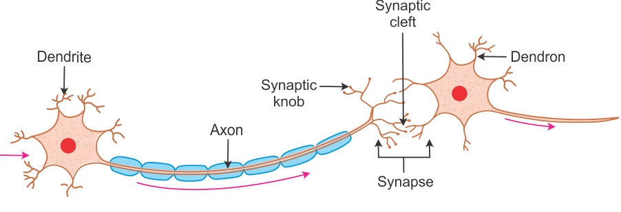
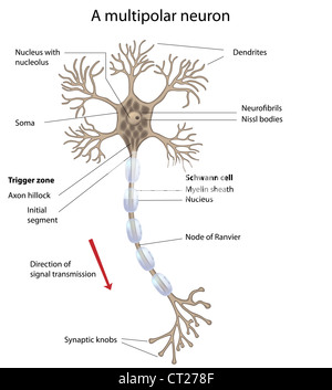
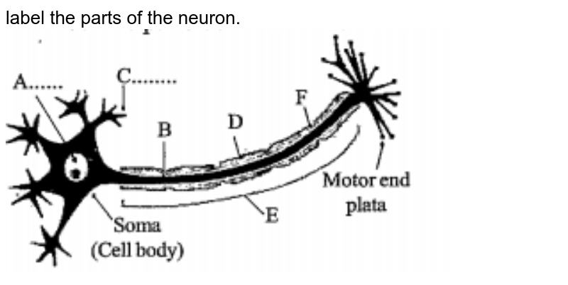
Schwann Cell Anatomy - Human Anatomy - GUWS Medical Figure 25.1 Label this diagram of a motor neuron. Figure 25.1 Label this diagram of a motor neuron. Figure 25.2 Label the features of the myelinated nerve fiber. Figure 25.3 Micrograph of a multipolar neuron and neuroglia from a spinal cord smear (100x micrograph enlarged to 600x).-Nerve fiber (axon) general name for processes (either dendrites ... Neuron under Microscope with Labeled Diagram - AnatomyLearner But, first, let's try to identify the following features from a neuron with the help of a labelled diagram. Cell body or perikaryon of a neuron Nucleus, cytoplasm, the plasma membrane of a neuron Nissl bodies in the cell body of a neuron An initial segment of axon and axon hillock Dendrites and axons of a neuron Axolemma and myelin sheath
en.wikipedia.org › wiki › AcetylcholinesteraseAcetylcholinesterase - Wikipedia The liberated choline is taken up again by the pre-synaptic neuron and ACh is synthesized by combining with acetyl-CoA through the action of choline acetyltransferase. [19] [20] A cholinomimetic drug disrupts this process by acting as a cholinergic neurotransmitter that is impervious to acetylcholinesterase's lysing action.

Diagram of neuron with labels
General Structure of a Neuron (Nerve Cell) | GetBodySmart General Structure of a Neuron (Nerve Cell) Start Quiz. Learn this topic from scratch or practice what you already know with these interactive spaced repetition-inspired anatomy quizzes. Learn anatomy faster and. remember everything you learn. Start Now. <. Neuron Diagram, Structure & Function | What Is a Neuron? - Video ... Neurons transmit signals between the brain and spinal cord to the rest of the body. They contain several unique and important structures that allow them to perform this important job. In the neuron... Important Question for Class 10 Science Control and Coordination PDF Sequence of events can be summarised as : Photoreceptors in eye → Sensory (Receptor) neuron → Brain → Motor (Effector) neuron → Eye muscle → Constriction of pupils. Question 7. (a) Draw a neat diagram of a neuron and label (i) dendrite and (ii) axon. (b) Which part of the human brain is: (i) the main thinking part of the brain?
Diagram of neuron with labels. drag the labels to their appropriate locations on the diagram of the ... Every neuron forms connections with other neurons. These connections are known as synapsis. During synapsis, when a presynaptic neuron sends information, it releases neurotransmitters. This event is done through exocytosis. The neurotransmitter is a molecule that travels through the synaptic space forward to the other neuron. Histology of neurons: Morphology and types of neurons | Kenhub Neurons have been grouped into two broad categories: those found in the central nervous system (brain and spinal cord) and those in the peripheral nervous system. In the central nervous system, they are found in clusters referred to as nuclei, or in layers also known as laminae. However, in the peripheral nervous system, they are found in ganglia. neuron | Definition & Functions | Britannica Bundles of fibres from neurons are held together by connective tissue and form nerves. Some nerves in large vertebrates are several feet long. A sensory neuron transmits impulses from a receptor, such as those in the eye or ear, to a more central location in the nervous system, such as the spinal cord or brain. Nervous system: Structure, function and diagram | Kenhub Neurons, or nerve cell, are the main structural and functional units of the nervous system. Every neuron consists of a body (soma) and a number of processes (neurites). The nerve cell body contains the cellular organelles and is where neural impulses ( action potentials) are generated.
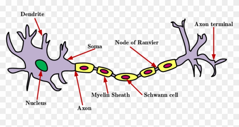
11.3: Neurons - Biology LibreTexts Figure 11.3. 2 shows the structure of a typical neuron. The main parts of a neuron are labeled in the figure and described below. Figure 11.3. 2: Somatic Motor Neuron with cell body, axon, axon, myelin sheath, nodes of Ranvier, axon terminal, dendrites, synaptic end of the bulbs, and other associated structures. Synaptic Cleft | Anatomy, Structure, Diseases & Functions Synaptic cleft is a space between two neurons, connecting them to one another forming a synapse. It is bound on one side by pre-synaptic neuron and. have post-synaptic neuron on the other side. The presynaptic neuron is always. an axon terminal. Depending on the type of synapse, the post-synaptic neuron. may be; Cells of the Nervous System - Neurons - TeachMePhysiology The nervous system comprises of two groups of cells, glial cells and neurones. Neurones are responsible for sensing change in their environment and communicating with other neurones via electrochemical signals. Glial cells work to support, nourish, insulate neurones and remove the waste products of metabolism. What Are Neurons? Draw A Clear And Labeled Diagram Of A Neuron. Doubt ... Draw a clear and labeled diagram of a neuron. 1 Answer. Muskan Anand. 2022-07-09T04:01:19.933000Z; A neuron is a specialized cell, primarily involved in transmitting information through electrical and chemical signals. They are found in the brain, spinal cord and the peripheral nerves. A neuron is also known as the nerve cell.
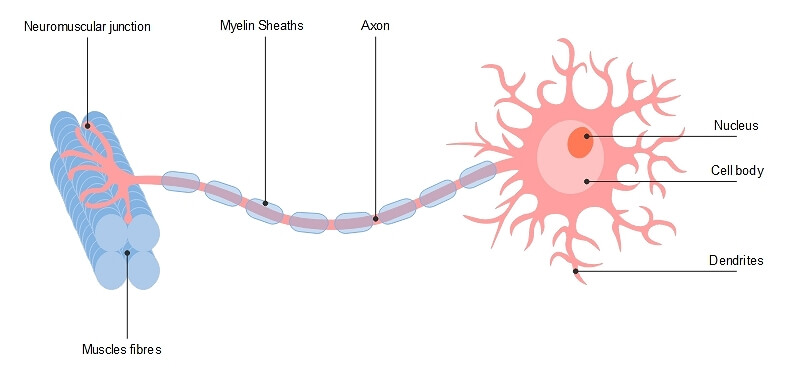
Free Nervous System Worksheets and Printables - Homeschool Giveaways Neuron Printable Clipart and Labeling Sheets - This is an amazing selection of neuron, or nerve cell, clip art for you and your kids to print and label. Inside Out Anatomy: The Brain - This Inside-Out worksheet shows the structure and function of the brain. Kids will color the different parts as they learn about this fascinating body system. Labeled Neuron Diagram| EdrawMax Template The following labeled diagram shows the parts of a neuron. In order to make it more understandable to the students, we have added all the functions of the Neuron in the labeled diagram. The major parts of the Neuron are Dendrites, Cell Body, Cell Membrane, Axon Hillock, Node of Ranvier, Schwann Cell, Axon Terminal, Myelin Sheath, Axon, and Nucleus. Types of Neurons: Parts, Structure, and Function - Verywell Health Different types of neurons include sensory, motor, and interneurons, as well as structurally-based neurons, which include unipolar, multipolar, bipolar, and pseudo-unipolar neurons. These cells coordinate bodily functions and movement so quickly, we don't even notice it happening. A Word From Verywell (Get Answer) - Label the parts of the neuron in the diagram. Not all ... The diagram below is of a nerve cell or neuron. Add the following labels to the diagram: Dendrites Muscle fibers Axon terminals Axon Myelin sheath Cell body 2. Color in the diagram as suggested below.
mccormickml.com › 2013/08/15 › radial-basis-functionRadial Basis Function Network (RBFN) Tutorial · Chris McCormick Aug 15, 2013 · The shape of the RBF neuron’s response is a bell curve, as illustrated in the network architecture diagram. The neuron’s response value is also called its “activation” value. The prototype vector is also often called the neuron’s “center”, since it’s the value at the center of the bell curve. The Output Nodes
What Is a Neuron? Diagrams, Types, Function, and More - Healthline These neurons allow the brain and spinal cord to communicate with muscles, organs, and glands all over the body. There are two types of motor neurons: lower and upper. Lower motor neurons carry...
Identify the parts labeled 1, 2, 3, 4, and 5 of the neuron. The 3rd number is Myelin sheath which is responsible for the increase in speed of the signals, 4th number is the Schwann cell that produces myelin sheath and the 5th number is Axon that transfer signals to other cells and organs so we can conclude that these are the label parts of neuron. Learn more: brainly.com/question/25200794 Advertisement
Neuron Structure Worksheet Answers - Png Becerra The diagram below is of a nerve cell or neurone. Sends messages from the cell body to the dendrites of other neurons. Add the following labels to the diagram. Start studying neuron structure worksheet. Students will use evidence from the video to answer questions and gain .
Multipolar Neurons - Structure and Functions | GetBodySmart Multipolar neurons have three or more processes attached to the cell bodies. 1. 2. One process serves as the axon, which conducts electrochemical impulses (action potentials) between cells. 1. 2. The remaining processes are dendrites. Togather, the cell body and dendrites form the receptive zone of multipolar neurons. 1.
41.11: Human Osmoregulatory and Excretory Systems - Nephron- The ... The renal corpuscle, located in the renal cortex, is composed of a network of capillaries known as the glomerulus, as well as a cup-shaped chamber that surrounds it: the glomerular or Bowman's capsule. The renal tubule is a long, convoluted structure that emerges from the glomerulus. It can be divided into three parts based on function.
Activation functions in Neural Networks - GeeksforGeeks Definition of activation function:- Activation function decides, whether a neuron should be activated or not by calculating weighted sum and further adding bias with it. The purpose of the activation function is to introduce non-linearity into the output of a neuron. Explanation :- We know, neural network has neurons that work in correspondence ...
Neurons: Meaning, Types, Functions, Diagrams - Embibe The main three parts of a neuron are the cell body or soma, axon, and dendrite. These parts are responsible for transmitting chemical and electrical signals. Cell body or Soma: The cell body or Soma is also called a cyton. It contains a nucleus and cytoplasm that connects to dendrites.
› ~kimscott › slides7. Artificial neural networks - Massachusetts Institute of ... Neuron Unit Synapse Connection Synaptic strength Weight Firing frequency Signals pass fromUnit output Table 1 (left): Corresponding terms from biological and artificial neural networks. Adapted from Adapted from Mehrotra, Mohan, & Ranka. Figure 1 (below): Schematic diagram of a standard neural network design. the input units
Neuron Diagram Labeled | EdrawMax Template It is an effective form of self-assessment, enabling students to check their understanding. In the following diagram, we have illustrated the important parts of the Neuron. In the following Neuron labeled diagram, we have dendrite, cell body, axon, myelin sheath, Schwann cell, a node of Ranvier, axon terminal, and nucleus.
› articles › s41593/022/01041-5Single-neuron projectome of mouse prefrontal cortex | Nature ... Mar 31, 2022 · Top, a diagram and an example neuron with the over-represented pattern of projections to MOp, SSp, and SSs. Extended Data Fig. 6 Diversity of projections to lateral and central cortical subnetworks.
CBSE Class 10 Science Important Biology Diagrams For Last Minute ... Important Biology diagrams for CBSE Class 10 Board Exam 2022 are given below: 1. Neuron. Neurons or the nerve cells form the basic components of the nervous system. A typical neuron possesses a ...
Neuron Labeling Worksheet - Nervous System Label The Neuron Label the parts of the neuron with the correct title: This is a diagram of a neuron. Click on the dot and type in the correct structure. Click on the template below to see it in its own window and . Use this video to label and color the neuron. Consider this main learning worksheet to teach all about the fabulous neuron cells and the 6 parts.
byjus.com › biology › skin-diagramSkin Diagram with Detailed Illustrations and Clear Labels - BYJUS Skin Diagram The largest organ in the human body is the skin, covering a total area of about 1.8 square meters. The skin is tasked with protecting our body from external elements as well as microbes.
Anatomy and Physiology Diagram | Quizlet Using Figure 7.1, identify the following: The metabolic center of the neuron is indicated by _____. A) Label D B) Label F C) Label A D) Label H
pressbooks.uwf.edu › medicalterminology › chapterNervous System – Medical Terminology for Healthcare Professions Labels read (top, left): pons, inferior olive, (top, right) cerebellum, deep cerebellar white matter (arbor vitae). In the top panel, a lateral view labels the location of the cerebellum and the deep cerebellar white matter. In the bottom panel, a photograph of a brain, with the cerebellum in pink is shown. [Return to Figure 8.7].
byjus.com › biology › diagram-of-neuronA Labelled Diagram Of Neuron with Detailed Explanations - BYJUS The diagram or the structure of the Neuron is useful for both Class 11 and 12 board exams as it has been repetitively asked in the board examinations. It is also one among the few topics having the highest weightage of marks. Learn More: Difference between Sensory and Motor Neuron. Diagram Of Neuron with Labels
Interneurons: Function, Diagram & Location - Study.com Relay interneurons, on the other hand, have long axons and can therefore complete circuits across larger regions of the body and brain. Interneuron Structure and Diagram All neurons, including...
Important Question for Class 10 Science Control and Coordination PDF Sequence of events can be summarised as : Photoreceptors in eye → Sensory (Receptor) neuron → Brain → Motor (Effector) neuron → Eye muscle → Constriction of pupils. Question 7. (a) Draw a neat diagram of a neuron and label (i) dendrite and (ii) axon. (b) Which part of the human brain is: (i) the main thinking part of the brain?
Neuron Diagram, Structure & Function | What Is a Neuron? - Video ... Neurons transmit signals between the brain and spinal cord to the rest of the body. They contain several unique and important structures that allow them to perform this important job. In the neuron...
General Structure of a Neuron (Nerve Cell) | GetBodySmart General Structure of a Neuron (Nerve Cell) Start Quiz. Learn this topic from scratch or practice what you already know with these interactive spaced repetition-inspired anatomy quizzes. Learn anatomy faster and. remember everything you learn. Start Now. <.



:max_bytes(150000):strip_icc()/purkinje_neuron-599da56d396e5a0011a0d344.jpg)












Post a Comment for "43 diagram of neuron with labels"