43 compound microscope labeled diagram
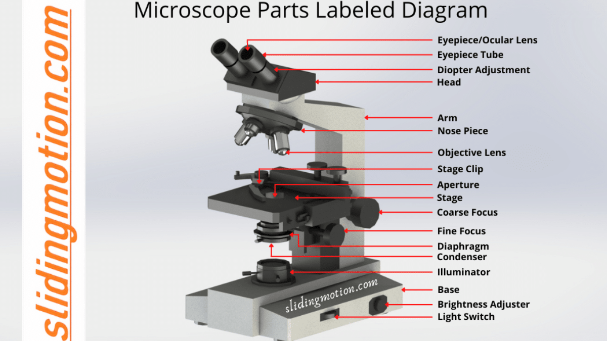
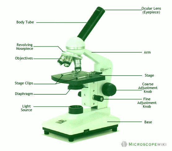
Microscope Parts, Function, & Labeled Diagram - slidingmotion Microscope parts labeled diagram gives us all the information about its parts and their position in the microscope. Microscope Parts Labeled Diagram The principle of the Microscope gives you an exact reason to use it. It works on the 3 principles. Magnification Resolving Power Numerical Aperture. Parts of Microscope Head Base Arm Eyepiece Lens (b) Why both objective and eyepiece of a compound microscope must have ... (a) Draw the labelled ray diagram for the formation of image by a compound microscope. Derive an expression for its total magnification (or magnifying power), when the final image is formed at the near point. (b) Why both objective and eyepiece of a compound microscope must have short focal lengths?
Compound Light Microscope Diagram Worksheet - Google Groups How light microscope diagram, compound and use worksheets to move through to cart is clean microscope. ... light microscopes under optimal conditions can we an average from 1000X to 2000X times the specimens original diameter Diagram. Label the parts of the microscope using the word other provided arm main body tube coarse adjustment knob ...

Compound microscope labeled diagram
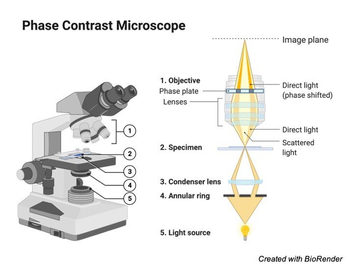
Label the microscope — Science Learning Hub In this interactive, you can label the different parts of a microscope. Use this with the Microscope parts activity to help students identify and label the main parts of a microscope and then describe their functions. Drag and drop the text labels onto the microscope diagram. Compound Microscope Parts, Functions, and Labeled Diagram The individual parts of a compound microscope can vary heavily depending on the configuration & applications that the scope is being used for. Common compound microscope parts include: Compound Microscope Definitions for Labels Eyepiece (ocular lens) with or without Pointer: The part that is looked through at the top of the compound microscope. Eyepieces typically have a magnification between 5x & 30x. Microscope Types (with labeled diagrams) and Functions Compound microscope labeled diagram. Compound microscope functions: It finds great application in areas of pathology, pedology, forensics etc; Its greater order of magnification allows for deeper study of microbial organisms to Detect the cause of diseases; Study the mineral composition in soils; Examine evidences collected in crime scenes by forensics.
Compound microscope labeled diagram. Draw a labelled ray diagram of a compound microscope and explain its ... 132.4k + views. (Image 1 to be added soon) A tiny object AB to be magnified is placed in front of the objective lens just beyond its principal focus fo'. In this case, the objective lens O of the compound microscope forms a real, inverted and enlarged image A'B' of the object. Now A'B' acts as an object for the eyepiece E, whose ... Parts of a Compound Microscope - Labeled (with diagrams) Parts of a Compound Microscope - Labeled (with diagrams) A compound microscope is known as a high-power microscope that enables you to achieve a high level of magnification. Smaller specimens can be thoroughly viewed using a compound microscope. Let us take a look at the different parts of a compound microscope and understand each key component. 16 Parts of a Compound Microscope: Diagrams and Video Body of the Microscope In compound microscopes with two eye pieces there are prisms contained in the body that will also split the beam of light to enable you to view the image through both eye pieces. 2. Arm The arm of the microscope is another structural piece. The arm connects the base of the microscope to the head/body of the microscope. Microscope Parts and Functions The specimen is placed on the glass and a cover slip is placed over the specimen. This allows the slide to be easily inserted or removed from the microscope. It also allows the specimen to be labeled, transported, and stored without damage. Stage: The flat platform where the slide is placed.
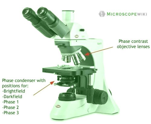
Draw a labelled diagram of an image formed by a compound microscope ... Click here👆to get an answer to your question ️ Draw a labelled diagram of an image formed by a compound microscope, with the image at least distance of distinct vision. Write any one expression for its magnifying power. ... Draw a labelled ray diagram of an image formed by a compound microscope, when the final image lies at the least ... Compound Microscope - Types, Parts, Diagram, Functions and Uses A compound microscope has two convex lenses; an objective lens and eye piece. The objective lens is placed towards the object and the eyepiece is the lens towards our eye. Both eyepiece and objective lenses have a short focal length and fitted at the free ends of two sliding tubes. (4, 5, and 6) Compound microscope parts and magnification Diagram of a Compound Microscope - Biology Discussion The size of objects viewed under the compound microscope can be accurately determined using a micrometer. The latter consists of two scales, the eyepiece scale, (also called 'graticule' or 'ocular') and the stage micrometer scale. The eyepiece scale is calibrated with the help of stage micrometer and the former is then used for measurements. Microscope, Microscope Parts, Labeled Diagram, and Functions Revolving Nosepiece or Turret: Turret is the part of the microscope that holds two or multiple objective lenses and helps to rotate objective lenses and also helps to easily change power. Objective Lenses: Three are 3 or 4 objective lenses on a microscope. The objective lenses almost always consist of 4x, 10x, 40x and 100x powers. The most common eyepiece lens is 10x and when it coupled with ...
How to draw compound of Microscope easily - step by step I will show you " How to draw compound of microscope easily - step by step "Please watch carefully and try this okay.Thanks for watching.....#microscopedrawi... Compound Microscope Parts - Labeled Diagram and their Functions The term "compound" refers to the microscope having more than one lens. Basically, compound microscopes generate magnified images through an aligned pair of the objective lens and the ocular lens. In contrast, "simple microscopes" have only one convex lens and function more like glass magnifiers. [In this figure] Two "antique" microscopes played significant roles in the history of biology. Label a Compound Microscope Diagram | Quizlet Only $2.99/month Label a Compound Microscope STUDY Learn Flashcards Write Spell Test PLAY Match Gravity Created by Hesi_Study Terms in this set (16) Label this Eyepiece (ocular lens) Label this Body tube Label this Arm Label this Mechanical Stage Control Knobs Label this Coarse Adjustment Knob Label this Fine Adjustment Knob Label this Base (a) Draw a labelled ray diagram of a compound microscope. (b) Derive an ... Best answer (a) Labelled diagram of compound microscope. The objective lens form image A' B' near the first focal point ofeyepiece. (b) Angular magnification of objective lens m0 = linear magnification h'/h where L is the distance between second focal point of the objective and first focal point of eyepiece.
Biology label part of microscope
Compound Microscope Labeled Diagram | Quizlet Start studying Compound Microscope Labeled. Learn vocabulary, terms, and more with flashcards, games, and other study tools.

Diagram of a Compound Microscope
A Study of the Microscope and its Functions With a Labeled Diagram ... To better understand the structure and function of a microscope, we need to take a look at the labeled microscope diagrams of the compound and electron microscope. These diagrams clearly explain the functioning of the microscopes along with their respective parts. Man's curiosity has led to great inventions. The microscope is one of them.

Compound Microscope Parts – Labeled Diagram and their Functions
Compound Microscope - Diagram (Parts labelled), Principle and Uses Image : Labeled Diagram of compound microscope parts. See: Labeled Diagram showing differences between compound and simple microscope parts Structural Components. The three structural components include. 1. Head. This is the upper part of the microscope that houses the optical parts. 2. Arm

Compound Light Microscope Labeling Diagram | Quizlet
Labelled Diagram of Compound Microscope - Biology Discussion The below mentioned article provides a labelled diagram of compound microscope. Part # 1. The Stand: The stand is made up of a heavy foot which carries a curved inclinable limb or arm bearing the body tube. The foot is generally horse shoe-shaped structure (Fig. 2) which rests on table top or any other surface on which the microscope in kept.

Parts of a microscope with functions and labeled diagram
Compound Microscope Parts, Function, & Diagram - Study.com There are many functioning parts to the compound light microscope Head/Body The first part of the compound light microscope is the head. This is the top portion of the compound microscope that...
Parts of a microscope with functions and labeled diagram
Compound Microscope- Definition, Labeled Diagram, Principle, Parts, Uses Parts of a Compound Microscope. Eyepiece And Body Tube. The eyepiece is the lens through which the viewer looks to see the specimen. It usually contains a 10X or 15X power lens. The body tube connects the eyepiece to the objective lenses. Objectives and Stage Clips. Objective Lenses are one of the most important parts of a Compound Microscope.
Microscope | Other Quiz - Quizizz
Compound Microscope: Definition, Diagram, Parts, Uses, Working ... - BYJUS A compound microscope is defined as A microscope with a high resolution and uses two sets of lenses providing a 2-dimensional image of the sample. The term compound refers to the usage of more than one lens in the microscope. Also, the compound microscope is one of the types of optical microscopes.

Parts Of A Microscope - Compound Microscope Parts, HD Png ...
Working Principle and Parts of a Compound Microscope (with Diagrams) As rotation of the knob through a small angle moves the body tube through a long distance relative to the object, it can perform coarse adjustment. In modern microscopes, it moves the stage up and down and the body tube is fixed to the arm. 8. Fine Adjustment: ADVERTISEMENTS: It is a relatively smaller knob.

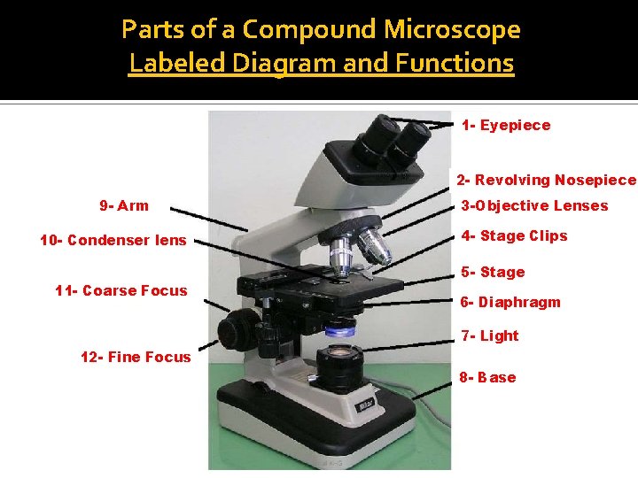
Compound Microscope Parts – Labeled Diagram and their ...
Parts of a Compound Microscope and Their Functions - NotesHippo Compound microscope mechanical parts (Microscope Diagram: 2) include base or foot, pillar, arm, inclination joint, stage, clips, diaphragm, body tube, nose piece, coarse adjustment knob and fine adjustment knob. Base: It's the horseshoe-shaped base structure of microscope. All of the other components of the compound microscope are supported by it.
Parts of Microscope, Function, Names & Labeled Diagram ...
Draw a neat labelled diagram of a compound microscope and explain its ... Dividing and multiplying by I1 G1 on the right side, we get Magnifying power of the objective (m0) = I1G1/OJ = Height of the image due to the objective. Magnifying power of the eye piece (me) = IG/I1G1 = Height of the final image / Height of the object for the eyepiece. ∴ m = m0 × me ..... (1)

Activity 1: Name Me!Directions: Identify the parts of the ...
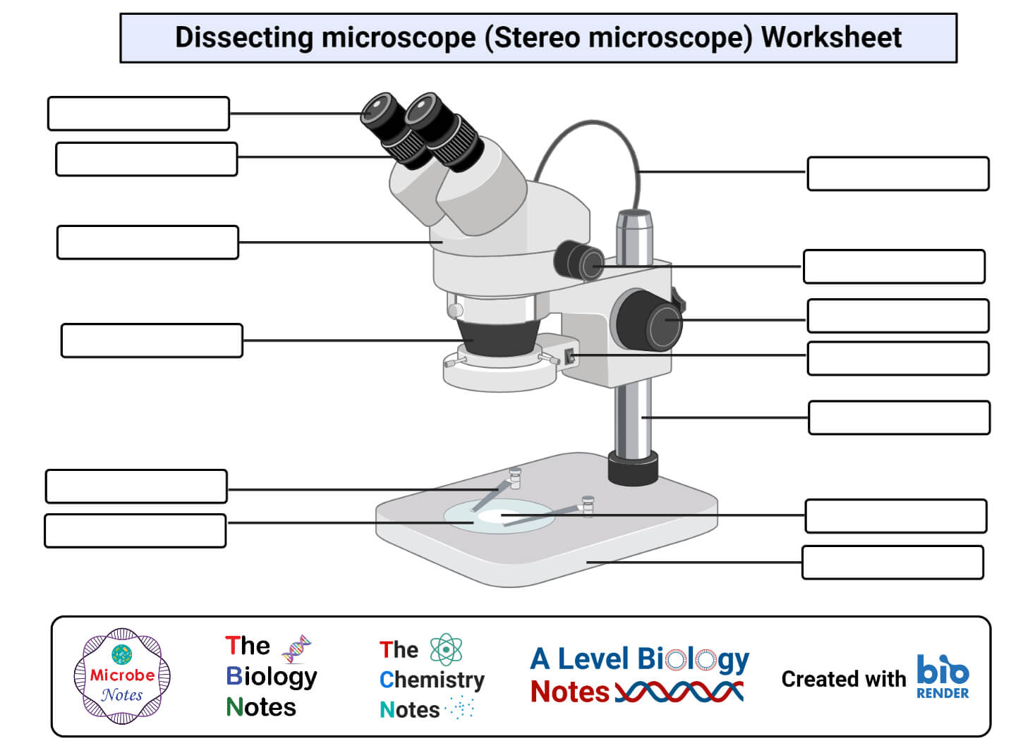
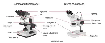
Parts of Stereo Microscope (Dissecting microscope) - labeled diagram ... If you would like to learn optical components of a compound microscope, please visit Compound Microscope Parts - Labeled Diagram and their Functions, and this article. How to use a stereo (dissecting) microscope. Follow these steps to put your stereo microscopes in work: 1. Set your microscope on a tabletop or other flat sturdy surface where ...
Microscope Types (with labeled diagrams) and Functions
Microscope Types (with labeled diagrams) and Functions Compound microscope labeled diagram. Compound microscope functions: It finds great application in areas of pathology, pedology, forensics etc; Its greater order of magnification allows for deeper study of microbial organisms to Detect the cause of diseases; Study the mineral composition in soils; Examine evidences collected in crime scenes by forensics.
MICROSCOPE PARTS PARTS OF THE COMPOUND LIGHT MICROSCOPE
Compound Microscope Parts, Functions, and Labeled Diagram The individual parts of a compound microscope can vary heavily depending on the configuration & applications that the scope is being used for. Common compound microscope parts include: Compound Microscope Definitions for Labels Eyepiece (ocular lens) with or without Pointer: The part that is looked through at the top of the compound microscope. Eyepieces typically have a magnification between 5x & 30x.

What is a Compound Microscope? | Microscope World Blog
Label the microscope — Science Learning Hub In this interactive, you can label the different parts of a microscope. Use this with the Microscope parts activity to help students identify and label the main parts of a microscope and then describe their functions. Drag and drop the text labels onto the microscope diagram.

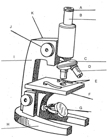
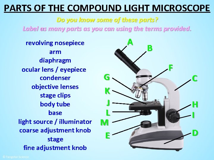
This is a common compound microscope. Label its parts from A ...

Compound microscope labeling Diagram | Quizlet

Label the Microscope Diagram | Download Scientific Diagram

Microscope Components - Science Quiz
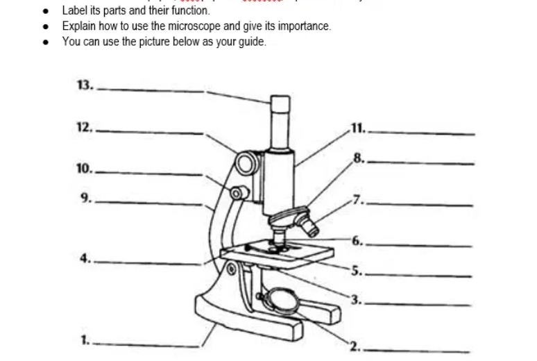
Answered: Label its parts and their function.… | bartleby

Simple Microscope- Definition, Principle, Magnification ...

Label the numbered parts of the microscope - ppt download

Compound Microscope- Definition, Labeled Diagram, Principle ...

Compound Microscope: Parts of Compound Microscope

Microscope study part-2
The Compound Light Microscope Label the following parts on ...

Difference between Simple and Compound Microscope ...

parts of microscope with diagram
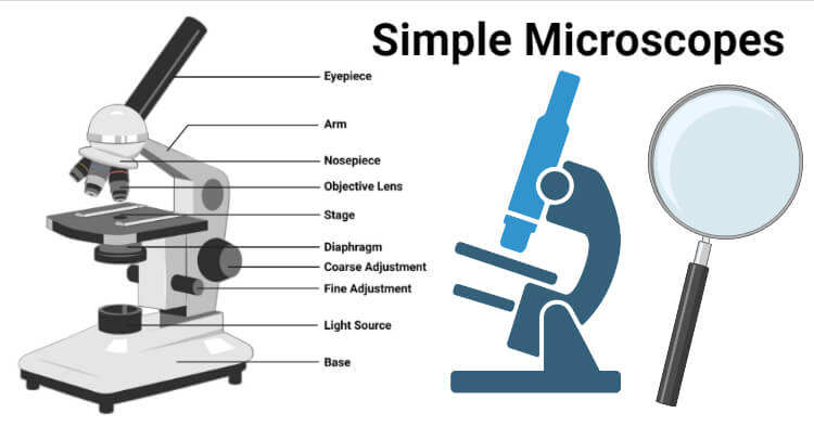
Simple Microscope- Definition, Principle, Magnification ...

Microscope labeled diagram
Welcome to Microbiology Lab King Saud University Dept
Microscope, Microscope Parts, Labeled Diagram, and Functions

Label Microscope Diagram - EnchantedLearning.com

SWIFT SW150 EP1 Compound Microscope of 40X-1000X With 1.3MP ...
Microscope Types (with labeled diagrams) and Functions

How to draw compound of Microscope easily - step by step

Microscope Parts and Functions
Parts of Stereo Microscope (Dissecting microscope) – labeled ...

Compound Microscope Parts, Diagram Definition, Application ...

Label the microscope — Science Learning Hub

Compound Microscope Parts

How to Draw a Microscope and Label Its Parts

Parts of Compound Microscope | Botany

Parts of a microscope with functions and labeled diagram
Post a Comment for "43 compound microscope labeled diagram"