38 correctly label the following anatomical features of the eye.
Parts of the Eye and Their Functions - Robertson Opt The iris is the area of the eye that contains the pigment which gives the eye its color. This area surrounds the pupil, and uses the dilator pupillae muscles to widen or close the pupil. This allows the eye to take in more or less light depending on how bright it is around you. If it is too bright, the iris will shrink the pupil so that they ... 8B4C53F7-CF59-48CF-BBD3-002197386B88.jpeg - Course Hero View Homework Help - 8B4C53F7-CF59-48CF-BBD3-002197386B88.jpeg from BIO 203 at Bunker Hill Community College. Correctly label the following anatomical features of the surface of the brain. Cerebral
Eye anatomy and function - AboutKidsHealth The anatomy of the eye The eye has many parts that must work together to produce clear vision: The sclera, or white part of the eye, protects the eyeball. The pupil, or black dot at the centre of the eye, is an opening through which light can enter the eye. The iris, or coloured part of the eye, surrounds the pupil.
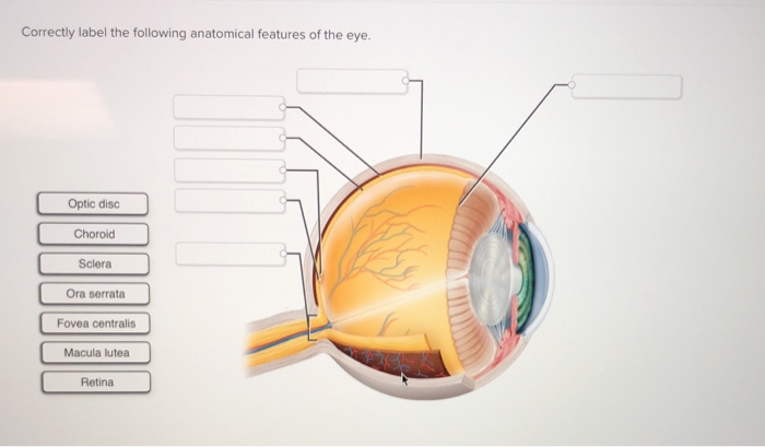
Correctly label the following anatomical features of the eye.
Solved Correctly label the following anatomical features of Transcribed image text: Correctly label the following anatomical features of the eye. Ora serrata Sclera Chorold Fovea centralis Optic disc Macula lutea Retina Correctly label the following anatomical features of the eye. Cornea Vitreous body Iris Pupil Ciliary body Suspensory ligaments Lens Previous question Next question AHCDW12Notes20.pdf - 20. Award: 10.00 points Problems?... The inner layer (tunica interna) consists of the retina and beginning of the optic nerve. The optical components of the eye are transparent elements that admit light rays, bend (refract) them, and focus images on the retina. They include the cornea, aqueous humor, lens, and vitreous body. The neural components are the retina and optic nerve. Senses Quiz Flashcards | Quizlet Correctly identify the following extrinsic muscles of the eyeball. Place the following labels in order indicating the passage of light through the eyeball. Start with the cornea at the top. 1. cornea 2. anterior chamber 3. pupil 4. posterior chamber 5. lens 6. vitreous humor 7. retina 8. choroid
Correctly label the following anatomical features of the eye.. The Structure and Anatomy of the Eye | SelectSpecs.com The eye consists of three layers of tissue which make up the wall of the eye. The sclera is the outermost layer of tissue, also called the white of the eye. This layer is a very stable fibrous membrane that continues to retain the shape of the eye and provides protection. Connected to the sclera are the extra-ocular or extrinsic muscles of the eye. Eye anatomy: A closer look at the parts of the eye Eye anatomy: A closer look at the parts of the eye. When surveyed about the five senses — sight, hearing, taste, smell and touch — people consistently report that their eyesight is the mode of perception they value (and fear losing) most. Despite this, many people don't have a good understanding of the anatomy of the eye, how vision works ... Correctly label the following anatomical features of the eye. Correctly ... Correctly label the following anatomical features of the eye. Cornea Vitreous body Pupil Ciliary body Lens Suspensory ligaments Iris The master budget of a small manufacturer would normally include all necessary component budgets except the capital... Posted 8 months ago Q: BYJUS BYJUS
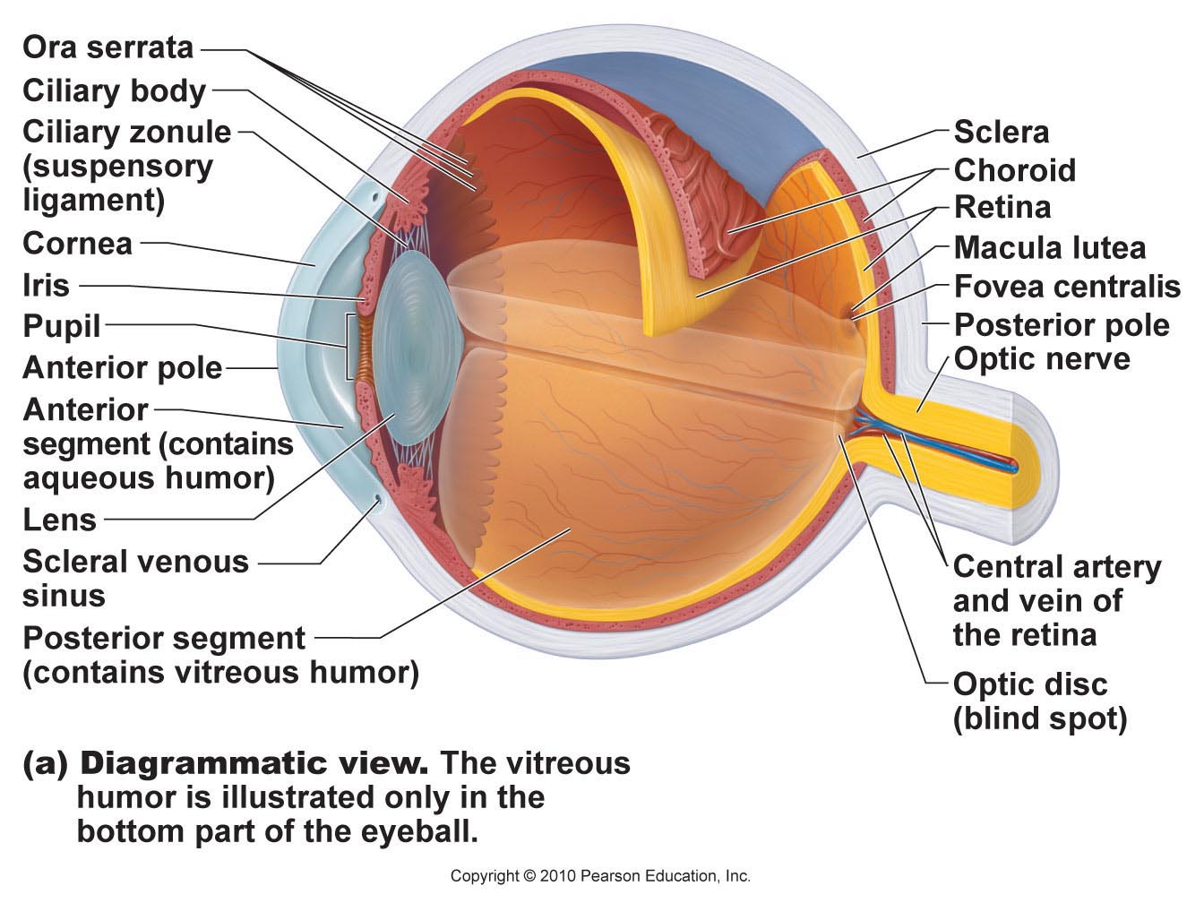
What Is the Path of Light Through the Eye? | Sciencing Updated April 29, 2018. By Jane Gingrich. The path of light through the eye begins with the objects viewed and how they produce, reflect or alter light in various ways. When your eyes receive light, it begins a second journey through the eye's optical parts that adjust and focus light to the nerves that carry images to your brain. Human Eye Anatomy - Parts of the Eye and Structure of the ... - Health Jade Structure of the Human Eye The eye is a hollow, spherical structure about 2.5 centimeters in diameter. Its wall has three distinct layers—an outer (fibrous) layer, a middle (vascular) layer, and an inner (nervous) layer. The spaces within the eye are filled with fluids that help maintain its shape. Figure 6. Structure of the human eye Connect Homework - Chapter 16 Flashcards | Quizlet Correctly label the anatomical elements of the projection pathways for pain. Place the following labels in order indicating the passage of light through the eyeball. Start with the cornea at the top. Correctly identify the following structures of the eye. Correctly label the anatomical elements of the tongue. Solved Correctly label the following anatomical features of - Chegg Question: Correctly label the following anatomical features of the cerebellum. Anterior lobe Anterior Vermis Folia Posterior lobe Posterior Cerebellar hemisphere (b) Superior view This problem has been solved! See the answer correctly label the following anatomical features of the cerebellum. Show transcribed image text Expert Answer
A&P 1 final Flashcards | Quizlet Left-handed people usually exhibit a wider frontal, parietal, and occipital lobe on the left compared to the same lobes on the right. Imagination, insight, and artistic skill are usually specialties of the right hemisphere. Lateralization develops with age and is not already developed at birth. Anatomy of the Eye | Kellogg Eye Center | Michigan Medicine How the Eye Works The five senses include sight, sound, taste, hearing and touch. Sight, like the other senses is closely related to other parts of our anatomy. The eye is connected to the brain and dependent upon the brain to interpret what we see. How we see depends upon the transfer of light. How the Eyes Work | National Eye Institute How the Eyes Work. All the different parts of your eyes work together to help you see. First, light passes through the cornea (the clear front layer of the eye). The cornea is shaped like a dome and bends light to help the eye focus. Some of this light enters the eye through an opening called the pupil (PYOO-pul). Parts of the Eye (Structure/Function) Quiz - Quizizz The part of your eye that gives it its color. It is a muscle that controls the size of your pupil. Q. The first part of the eye that light hits. Q. Controls how much light enters the eye by changes the size of the pupil. It is also the color part of your eye. Quiz not found!
Eye Anatomy Labeling - Anatomy Drawing Diagram
Solved Correctly label the following anatomical features of - Chegg Correctly label the following anatomical features of the eye. Optic disc Choroid Sclera Ora serrata Fovea centralis Macula lutea Retina Question: Correctly label the following anatomical features of the eye. Optic disc Choroid Sclera Ora serrata Fovea centralis Macula lutea Retina This problem has been solved! See the answer
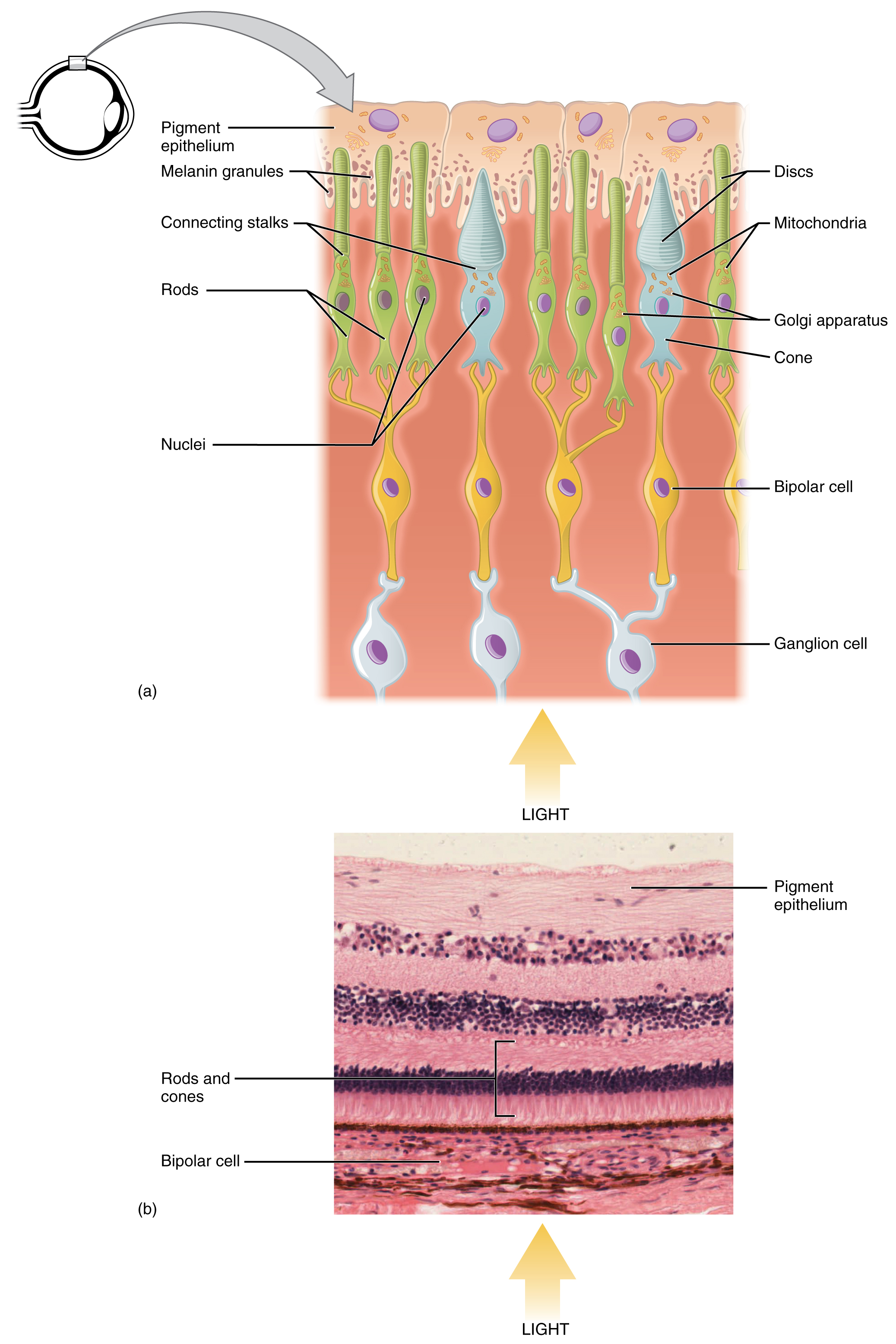
The top panel shows the cellular structure of the different cells in ...
15.5 Vision - Anatomy & Physiology 15.5 Vision Vision. Vision is the special sense of sight that is based on the transduction of light stimuli received through the eyes. The eyes are located within either orbit in the skull. The bony orbits surround the eyeballs, protecting them and anchoring the soft tissues of the eye (Figure 15.5.1).The eyelids, with lashes at their leading edges, help to protect the eye from abrasions by ...

Senses flashcards | Quizlet
AHCDW5SOL16.pdf - 16. Award: 10.00 points Problems ... - Course Hero Correctly label the following anatomical features of the elbow joint. Explanation: The elbow is a hinge with three joints: (1) the humeroulnar joint where the trochlear notch of the ulna encircles the trochlea of the humerus; (2) the humeroradial joint where the discshaped head of the radius meets the capitulum of the humerus; and (3) the ...
Believing Is Seeing | Steve Stern's Fieldnotes from a Random Walk ...
Free Science Flashcards about ANP1040 Exam 4 - StudyStack ANP1040 Exam 4. Correctly label the following anatomical features of a neuron. Correctly label the structures, areas, and concentrations associated with a cell's electrical charge difference across its membrane. ___ division carries signals to the smooth muscle in the large intestine.

30 Run For Cover Label - Online Labels Ideas
Anatomy Midterm Lecture Flashcards - Quizlet Correctly label the following anatomical features of the heart and thoracic cage. Correctly label the following structures related to the position of the heart in the thorax. Correctly label the following parts of the pericardium and the heart walls

35 Correctly Label The Following Functional Regions Of The Cerebral ...
Solved Correctly label the following anatomical features of - Chegg Correctly label the following anatomical features of the eye. Cornea Vitreous body Pupil Ciliary body Lens Suspensory ligaments Iris Question: Correctly label the following anatomical features of the eye. Cornea Vitreous body Pupil Ciliary body Lens Suspensory ligaments Iris This problem has been solved! See the answer Show transcribed image text

Spinal Cord Cross Section Diagram Spinal Cord Cross Section Diagram ...
BIOL3310 Lecture Exam Final Flashcards & Practice Test - Quizlet Correctly label the following anatomical features of the eye. In the _ division of the ANS the preganglionic nerve fibers are short, while in the _ division the preganglionic fibers are relatively long. Sympathetic; Parasympathetic Read each description below and determine whether it pertains to the medulla oblongata, the pons, or the midbrain.

Education and Resources | Retina Consultants of Western New York
Anatomy, Physiology & Pathology of the Human Eye - TedMontgomery.com eye structures. Several structures compose the human eye. Among the most important anatomical components are the conjunctiva, cornea, crystalline lens, extraocular muscles, iris, macula, optic nerve, retina, and vitreous humor. You can click on the names of each of these 9 ocular structures on the left (either in the upper picture or in the ...

Print Anatomy Exam 2 flashcards | Easy Notecards
human physiology lab manual answers Best Way To Study Human Anatomy And Physiology - Master A&P In 3 Days . anatomy physiology human questions test study practice answers lab manual master mastering way. 33 Correctly Label The Following Anatomical Features Of The Elbow Joint ambitiousmares.blogspot.com. label following correctly anatomical features joint elbow solved
Post a Comment for "38 correctly label the following anatomical features of the eye."